



Ligaments of the uterus & ovary anatomy
Also known as: broad ligament, round ligament, cardinal ligament, uterosacral ligament, suspensory ligament of ovary, pelvic ligaments
The peritoneal folds and fibromuscular bands that suspend the uterus, tubes and ovaries: the broad ligament with its mesosalpinx and mesovarium, the round, cardinal and uterosacral ligaments, the suspensory and ovarian ligaments and the vesico-uterine pouch.
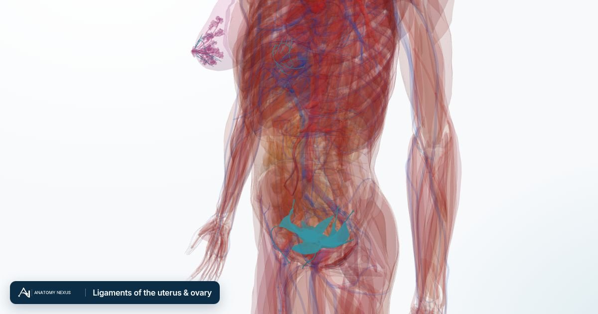
Structures in the atlas 18
The modelled pieces that make up the ligaments of the uterus & ovary in the 3D atlas. Each opens in the explorer.
- Broad ligament of uterus
- Left cardinal ligament
- Left mesosalpinx
- Left mesovarium
- Left ovarian ligament
- Left round ligament of uterus
- Left suspensory ligament of ovary
- Left uterosacral ligament
- Right cardinal ligament
- Right mesosalpinx
- Right mesovarium
- Right ovarian ligament
- Right round ligament of uterus
- Right suspensory ligament of ovary
- Right uterosacral ligament
- Suspensory ligaments of left breast
- Suspensory ligaments of right breast
- Vesico-uterine pouch
The female reproductive in brief
This layer brings the female body into an atlas whose reference model is male. Its pieces come from the united female model of the Human Reference Atlas (HRA), the reference built by the HuBMAP Consortium and the NIH from the Visible Human Female, scaled and placed so that the pubic symphysis, sacral promontory and hip bones of the female pelvis coincide with those of the BodyParts3D pelvis; the breasts sit on the reference chest wall at the level of the fourth to fifth rib. The uterus lies behind and above the bladder and in front of the rectum, tilted forward; the cervix opens into the top of the vagina, which runs down and back to the perineum; the uterine tubes (fallopian tubes) arch from the upper corners of the uterus out over the ovaries, which lie against the side walls of the pelvis; and the broad, round, cardinal and uterosacral ligaments and the suspensory and ovarian ligaments hold everything in place. The female bladder from the same model, with its trigone and neck, replaces the male bladder while the layer is shown, and a short schematic urethra runs from its neck down the front of the vagina to the vestibule (the HRA model has no urethra mesh). Each breast is shown as its body (fat and gland), the lobes of the mammary gland, the lactiferous ducts and sinuses, the suspensory ligaments, the areola with its tubercles and the nipple. Two things to know when reading it. The Visible Human Female was a woman of 59, and the whole layer is scaled to the male pelvis, so the uterus and ovaries are those of a woman past the menopause and smaller than the textbook figures on the organ pages. And because the reference skin is male, the breasts stand outside the skin overlay when both are shown. Switching this layer on hides the male reproductive organs, bladder and urethra so the two bodies are not drawn through each other; switching it off brings them back. The Female body preset shows the whole body with the female organs. The vulva and clitoris are not part of the HRA model and remain a written page.