



Breast Anatomy
Also known as: breast, mammary gland, mammary glands, nipple, chest
The breasts are paired modified sweat glands on the front of the chest wall, lying over the pectoralis major muscles and made of milk-producing lobes embedded in fat and tethered by fibrous ligaments. They grow under oestrogen and progesterone, make milk under prolactin and release it under oxytocin, and they are the site of the commonest cancer in women. The 3D model shows each breast from the NIH Human Reference Atlas on the reference chest wall: the body of the breast, the lobes of the mammary gland, the lactiferous ducts and sinuses, the suspensory ligaments, the areola with its tubercles and the nipple.
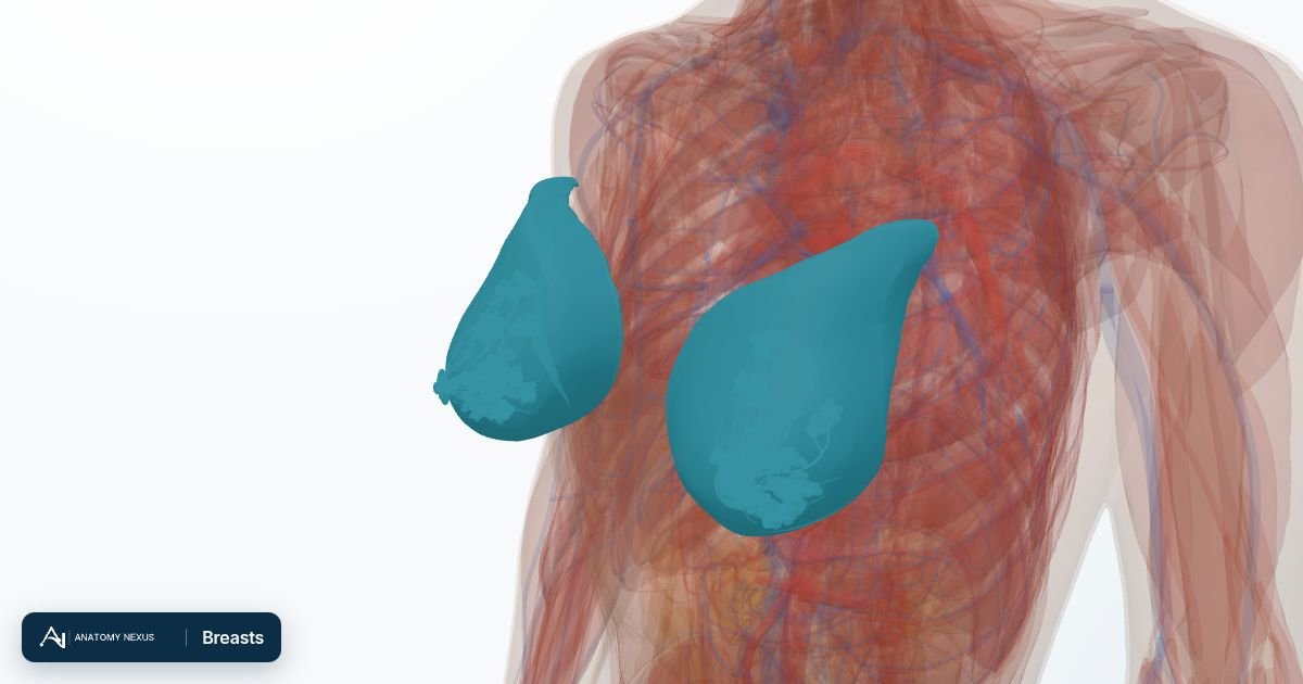
Key facts
| Location | On the front of the chest from the 2nd to the 6th rib and from the edge of the sternum to the mid-axillary line, with the axillary tail extending into the armpit |
|---|---|
| Glandular tissue | 15 to 20 lobes of branching tubulo-alveolar glands, each draining through a lactiferous duct; about 5 to 10 ducts open on the nipple and carry most of the milk |
| Support | Fat, and the suspensory ligaments (of Cooper) that run from the skin through the gland to the deep fascia over pectoralis major |
| Blood supply | Perforating branches of the internal thoracic artery, the lateral thoracic artery from the axillary artery, and perforating branches of the intercostal arteries |
| Lymph drainage | About three-quarters to the axillary nodes, most of the rest to the parasternal (internal mammary) nodes along the internal thoracic vessels |
| Nerve supply | Anterior and lateral cutaneous branches of the 4th to 6th intercostal nerves; the nipple lies in the T4 dermatome |
| Hormonal control | Oestrogen grows the ducts and fat, progesterone the lobules; prolactin drives milk production and oxytocin milk ejection |
Location and relations
Each breast lies in the superficial fascia of the front of the chest wall, extending from about the second to the sixth rib and from the lateral edge of the sternum to the mid-axillary line. A tongue of glandular tissue, the axillary tail, runs from the upper outer quadrant along the lower border of pectoralis major towards the armpit, which is why breast tissue and breast lumps can be found in the axilla. About two-thirds of the base rests on the fascia over pectoralis major and the rest on serratus anterior. Between the gland and the pectoral fascia lies the retromammary space, a layer of loose connective tissue that lets the breast move freely over the muscle; a cancer that grows through it fixes the breast to the chest wall. The nipple usually sits at the fourth intercostal space, though its position varies with breast size and age.
Structure: lobes, ducts and the nipple
The gland consists of 15 to 20 lobes arranged like the spokes of a wheel around the nipple, each a tubulo-alveolar gland whose lobules of secretory alveoli drain into progressively larger ducts. Every lobe empties through a lactiferous duct that runs towards the nipple, branching and lying close under the skin beneath the areola, where the classical description placed a widened lactiferous sinus; ultrasound of the lactating breast has not confirmed discrete reservoirs there, and milk is held in the alveoli and small ducts until the milk-ejection reflex moves it to the nipple, where about 5 to 10 openings carry most of it. Smooth muscle in the nipple makes it erect with cold, touch or suckling, and the pigmented areola carries the small bumps of the areolar (Montgomery) glands, whose oily secretion protects the skin during breastfeeding. The lobes are embedded in fat, which gives the breast most of its size and shape, and the gland is tethered by the suspensory ligaments of Cooper, fibrous bands running from the skin to the deep fascia. A tumour that shortens these ligaments draws in the skin above it, the dimpling that warns of cancer.
Blood supply and lymph
Blood reaches the breast from several directions: perforating branches of the internal thoracic (internal mammary) artery pierce the upper intercostal spaces beside the sternum to supply the medial part, the lateral thoracic artery from the axillary artery supplies the upper and lateral parts, and perforating branches of the posterior intercostal arteries arrive from behind. Veins drain mainly to the axillary and internal thoracic veins. Lymph drainage matters most clinically, because breast cancer spreads along it. About three-quarters of the lymph, especially from the lateral quadrants, passes to the axillary nodes, which surgeons group into three levels around pectoralis minor. Most of the rest drains to the parasternal (internal mammary) nodes along the internal thoracic vessels. Injecting a tracer beside a tumour identifies the first, or sentinel, node it drains to, which can be examined instead of clearing the whole axilla.
Innervation
Sensation from the skin of the breast is carried by the anterior and lateral cutaneous branches of the fourth, fifth and sixth intercostal nerves, with the supraclavicular nerves supplying the skin of the upper part. The nipple and areola lie in the fourth thoracic dermatome and are supplied chiefly by the lateral cutaneous branch of the fourth intercostal nerve, so nipple sensation can be reduced or lost if that branch is divided during breast surgery. These nerves also carry sympathetic fibres to the blood vessels and to the smooth muscle of the nipple; the gland itself has no motor supply, because milk secretion is controlled by hormones. Suckling stimulates sensory endings in the nipple and the signal travels to the hypothalamus, releasing prolactin and oxytocin. Surgery in the axilla can injure the intercostobrachial nerve, leaving numbness on the inner side of the upper arm.
Function and hormonal control
The purpose of the breast is lactation. During pregnancy, oestrogen, progesterone and placental hormones make the ducts branch and the alveoli multiply, and prolactin from the anterior pituitary rises steadily, but the high sex hormone levels block its action so that little milk is made before birth. Once the placenta is delivered, oestrogen and progesterone fall sharply and prolactin switches on full milk secretion within a few days; the first milk is colostrum, thick and rich in protein and antibodies. Suckling keeps the system going: input from the nipple sustains prolactin release and triggers oxytocin from the posterior pituitary, which contracts the myoepithelial cells around the alveoli and squeezes milk into the ducts, the let-down reflex. Supply follows demand, and the high prolactin of frequent breastfeeding often delays the return of ovulation and periods, though ovulation can return before the first period, so breastfeeding only prevents pregnancy reliably under the strict conditions of the lactational amenorrhoea method: fully breastfeeding, no periods yet returned and the baby under six months.
Changes across life
Before puberty, and in men throughout life, the breast is a small system of rudimentary ducts beneath the nipple that hormonal change or some medicines can enlarge (gynaecomastia). At puberty oestrogen grows the ducts and lays down fat, and progesterone develops the lobules once cycles become ovulatory; the lobules swell before each period, causing fullness and tenderness. Pregnancy enlarges the breast, and after the menopause glandular tissue gives way to fat. In trans women, breast growth usually begins about three to six months after starting oestrogen and continues over about two to three years, usually to a smaller size than in cisgender women. Trans women and non-binary people who have taken feminising hormones for several years are eligible for breast screening: those registered as female with a GP are invited in the same way as anyone else on the register, while those registered as male are not invited automatically and can ask their GP or local screening service to arrange it. Trans men who have not had chest surgery are eligible on the same basis and may also need to ask.
Clinical relevance
Breast cancer is the commonest cancer in women worldwide. It usually starts in the cells lining the ducts or lobules and can spread along the lymphatics to the axillary nodes, so a new lump, dimpled skin, a newly inverted nipple, blood-stained discharge or a lump in the armpit should be assessed promptly; a breast clinic combines examination, imaging and a needle biopsy. In England the NHS breast screening programme invites women, and anyone else registered female with a GP who has breast tissue, from the age of 50 up to their 71st birthday for mammography every three years. Most lumps are benign: fibroadenomas, cysts and fibrocystic change. A blocked duct during breastfeeding can lead to mastitis and, untreated, to an abscess. Breast cancer also occurs in men, though rarely. Chest masculinisation removes most but not all of the breast tissue, so a new lump still needs assessment, and augmentation implants sit over or under pectoralis major.
Structures in the atlas 16
The modelled pieces that make up the breasts in the 3D atlas. Each opens in the explorer.
- Areolar tubercles of left breast
- Areolar tubercles of right breast
- Body of left breast
- Body of right breast
- Lactiferous ducts of left breast
- Lactiferous ducts of right breast
- Lactiferous sinuses of left breast
- Lactiferous sinuses of right breast
- Left areola
- Left nipple
- Lobes of left mammary gland
- Lobes of right mammary gland
- Right areola
- Right nipple
- Suspensory ligaments of left breast
- Suspensory ligaments of right breast
The female reproductive in brief
This layer brings the female body into an atlas whose reference model is male. Its pieces come from the united female model of the Human Reference Atlas (HRA), the reference built by the HuBMAP Consortium and the NIH from the Visible Human Female, scaled and placed so that the pubic symphysis, sacral promontory and hip bones of the female pelvis coincide with those of the BodyParts3D pelvis; the breasts sit on the reference chest wall at the level of the fourth to fifth rib. The uterus lies behind and above the bladder and in front of the rectum, tilted forward; the cervix opens into the top of the vagina, which runs down and back to the perineum; the uterine tubes (fallopian tubes) arch from the upper corners of the uterus out over the ovaries, which lie against the side walls of the pelvis; and the broad, round, cardinal and uterosacral ligaments and the suspensory and ovarian ligaments hold everything in place. The female bladder from the same model, with its trigone and neck, replaces the male bladder while the layer is shown, and a short schematic urethra runs from its neck down the front of the vagina to the vestibule (the HRA model has no urethra mesh). Each breast is shown as its body (fat and gland), the lobes of the mammary gland, the lactiferous ducts and sinuses, the suspensory ligaments, the areola with its tubercles and the nipple. Two things to know when reading it. The Visible Human Female was a woman of 59, and the whole layer is scaled to the male pelvis, so the uterus and ovaries are those of a woman past the menopause and smaller than the textbook figures on the organ pages. And because the reference skin is male, the breasts stand outside the skin overlay when both are shown. Switching this layer on hides the male reproductive organs, bladder and urethra so the two bodies are not drawn through each other; switching it off brings them back. The Female body preset shows the whole body with the female organs. The vulva and clitoris are not part of the HRA model and remain a written page.