



Vulva & Clitoris Anatomy
Also known as: clitoris, labia, external genitalia, vestibule, mons pubis
The vulva is the external genitalia: the mons pubis, the labia majora and minora, the clitoris, the vestibule into which the urethra and vagina open, and the glands around them, resting on the front of the perineum between the thighs. Its centrepiece, the clitoris, is a densely innervated erectile organ homologous with the penis, most of which lies beneath the surface.
Key facts
| Parts | Mons pubis, labia majora, labia minora, clitoris, vestibule, Bartholin's and Skene's glands |
|---|---|
| Clitoris | Glans, body, two crura and two vestibular bulbs of erectile tissue, most of it beneath the surface |
| Openings | Urethral meatus in front and vaginal opening behind, both within the vestibule |
| Blood supply | Internal pudendal artery, with external pudendal branches from the femoral artery to the mons and front of the labia |
| Nerve supply | Pudendal nerve (S2-S4), with ilioinguinal and genitofemoral branches to the mons and anterior labia |
| Lymph | Superficial inguinal nodes in the groin |
| Development | From the same genital tubercle, folds and swellings that form the penis and scrotum |
Location and relations
The vulva occupies the front of the perineum, the diamond-shaped region between the thighs bounded by the pubic symphysis in front, the ischial tuberosities at the sides and the coccyx behind; the urogenital triangle in front holds the vulva, the anal triangle behind holds the anal canal and its external sphincter. The mons pubis is the fat pad over the symphysis, and the labia extend back from it to the perineal body, the fibromuscular knot between the vaginal opening and the anus into which the pelvic floor and perineal muscles insert. Beneath the skin lie the crura of the clitoris on the ischiopubic rami and the vestibular bulbs beside the vaginal opening, covered by the ischiocavernosus and bulbospongiosus muscles. The short female urethra, about 4 cm long, opens into the vestibule just in front of the vagina, one reason urinary tract infections are common in women.
Structure
The labia majora are two hair-bearing folds of skin and fat, meeting in front at the mons and behind near the perineal body. Within them lie the labia minora, thin hairless folds rich in sebaceous glands, vessels and nerve endings; in front each splits to form the prepuce (hood) over the glans of the clitoris and the frenulum beneath it, and behind they join at the fourchette. The clitoris has a glans, a short body, two crura along the ischiopubic rami and two vestibular bulbs flanking the vaginal opening, all erectile tissue that swells with blood during arousal; only the glans is visible, and most of the organ lies beneath the surface. The vestibule between the labia minora holds the urethral meatus, the vaginal opening, the ducts of Skene's (paraurethral) glands beside the urethra and the openings of Bartholin's (greater vestibular) glands on either side of the lower part of the vaginal opening.
Blood supply and lymph
Most of the vulva is supplied by the internal pudendal artery, a branch of the internal iliac that reaches the perineum by passing round the ischial spine and gives the perineal and posterior labial arteries, the artery of the bulb and the deep and dorsal arteries of the clitoris. The mons and the front of the labia receive external pudendal branches from the femoral artery. Venous blood returns through the internal pudendal veins and, from the clitoris, through the deep dorsal vein into the venous plexus around the bladder. The vestibular bulbs are so vascular that a straddle injury or a difficult birth can produce a large vulval haematoma. Lymph drains to the superficial inguinal nodes in the groin and then to the deep inguinal and external iliac nodes, which is why vulval cancer is staged by assessing the groin nodes and a vulval infection causes tender groin glands.
Innervation
The pudendal nerve (S2-S4) supplies most of the vulva. It gives perineal branches carrying the posterior labial nerves to the labia and vestibule, and the dorsal nerve of the clitoris, which makes the glans densely innervated and the most sensitive part of the genitalia. The mons and the front of the labia majora are supplied by the ilioinguinal nerve and the genital branch of the genitofemoral nerve (L1-L2), the back of the labia by the perineal branch of the posterior femoral cutaneous nerve. Autonomic fibres from the uterovaginal plexus reach the erectile tissue through the cavernous nerves and produce engorgement during arousal. A pudendal nerve block at the ischial spine anaesthetises the vulva and lower vagina for an instrumental birth or the repair of a tear, and in phalloplasty the dorsal nerve of the clitoris is joined to the nerve of the flap to give the neophallus sensation.
Function and development
The vulva protects the openings of the urethra and vagina and carries most of the sensation of the female genitalia. In arousal the clitoris and bulbs fill with blood, the labia swell and darken and the Bartholin's glands secrete mucus; the clitoris has no part in urination or reproduction, and its role is sensation and arousal. In the embryo the external genitalia of both sexes begin as the same genital tubercle, urogenital folds and labioscrotal swellings: without androgens the tubercle becomes the clitoris, the folds the labia minora and the swellings the labia majora, whereas testosterone and dihydrotestosterone turn the same tissues into the penis, penile urethra and scrotum, and the Bartholin's and Skene's glands correspond to the bulbourethral glands and prostate. These homologies underlie gender-affirming surgery: vaginoplasty fashions the glans of the penis into a neoclitoris, and metoidioplasty releases the enlarged clitoris to form a small phallus.
Changes across life
In childhood the labia majora are thin and hairless and the labia minora small. At puberty adrenal androgens bring pubic hair, and oestrogen deposits fat in the mons and labia majora, enlarges and darkens the labia minora and activates the glands. Pregnancy increases the blood supply, and vaginal birth stretches the vulva and perineum, where tears and episiotomies heal. After the menopause the loss of oestrogen thins the skin of the labia and vestibule, reduces labial fat and pubic hair, and causes dryness, itching and soreness: the genitourinary syndrome of menopause, which vaginal oestrogen relieves. Testosterone therapy in trans men and non-binary people enlarges the clitoris over months, typically to a few centimetres, a change that metoidioplasty makes use of. In trans women and some non-binary people, vaginoplasty constructs the labia from scrotal and penile skin and a neoclitoris from the glans of the penis, keeping its nerves and vessels.
Clinical relevance
A blocked Bartholin's gland forms a cyst beside the lower part of the vaginal opening; a small one often causes no symptoms, while a larger one feels uncomfortable, and an infected gland forms an abscess that is very painful and may need draining. Lichen sclerosus, a chronic inflammatory skin condition, causes itch, white thinned skin and scarring; it is treated with steroid ointment and is kept under review, because a small number of people with it go on to develop vulval cancer, which is rare and presents as a persistent lump, ulcer, itch or bleeding needing examination. Vulvodynia is persistent vulval pain with no visible cause; thrush, herpes and genital warts affect the vulval skin and are managed in sexual health clinics. Female genital mutilation, the cutting or removal of vulval tissue for non-medical reasons, is illegal in the UK and causes pain, scarring, urinary and menstrual problems, difficult childbirth and lasting psychological harm; specialist clinics offer support and deinfibulation where the opening has been narrowed. Wide variation in the size and shape of the labia is normal.
In the 3D atlas
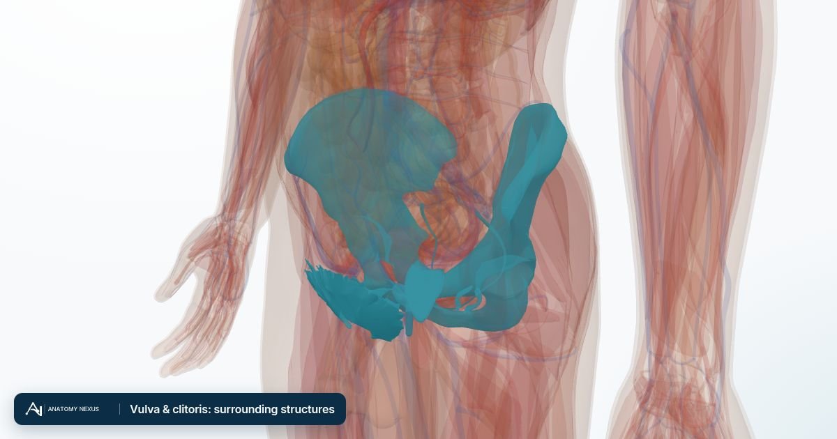
- External anal sphincter · 4 pieces
- Left hip bone
- Left internal pudendal vein
- Pubic hair
- Right hip bone
- Right internal pudendal vein
- Urethra (female, schematic)
- Vagina
The female reproductive in brief
This layer brings the female body into an atlas whose reference model is male. Its pieces come from the united female model of the Human Reference Atlas (HRA), the reference built by the HuBMAP Consortium and the NIH from the Visible Human Female, scaled and placed so that the pubic symphysis, sacral promontory and hip bones of the female pelvis coincide with those of the BodyParts3D pelvis; the breasts sit on the reference chest wall at the level of the fourth to fifth rib. The uterus lies behind and above the bladder and in front of the rectum, tilted forward; the cervix opens into the top of the vagina, which runs down and back to the perineum; the uterine tubes (fallopian tubes) arch from the upper corners of the uterus out over the ovaries, which lie against the side walls of the pelvis; and the broad, round, cardinal and uterosacral ligaments and the suspensory and ovarian ligaments hold everything in place. The female bladder from the same model, with its trigone and neck, replaces the male bladder while the layer is shown, and a short schematic urethra runs from its neck down the front of the vagina to the vestibule (the HRA model has no urethra mesh). Each breast is shown as its body (fat and gland), the lobes of the mammary gland, the lactiferous ducts and sinuses, the suspensory ligaments, the areola with its tubercles and the nipple. Two things to know when reading it. The Visible Human Female was a woman of 59, and the whole layer is scaled to the male pelvis, so the uterus and ovaries are those of a woman past the menopause and smaller than the textbook figures on the organ pages. And because the reference skin is male, the breasts stand outside the skin overlay when both are shown. Switching this layer on hides the male reproductive organs, bladder and urethra so the two bodies are not drawn through each other; switching it off brings them back. The Female body preset shows the whole body with the female organs. The vulva and clitoris are not part of the HRA model and remain a written page.