



Ovary & Fallopian Tube Anatomy
Also known as: ovary, fallopian tube, uterine tube, oviduct, adnexa
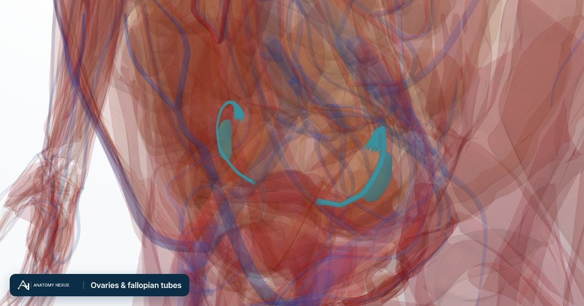
The ovaries are two almond-shaped glands about 3 cm long that lie against the side walls of the pelvis. Each holds a lifetime's supply of eggs, releases one at ovulation in most cycles and makes oestrogen and progesterone, while the fallopian tubes catch the egg, provide the site of fertilisation and carry the embryo to the uterus. The 3D model shows the ovaries and each uterine (fallopian) tube from the NIH Human Reference Atlas, its isthmus, ampulla and infundibulum with the fimbriae that fringe it (the short intramural part inside the uterine wall is not modelled separately), in place beside the uterus and against the pelvic side walls. The source body was a woman of 59 scaled to the reference pelvis, so the ovaries are smaller than the figure above.
Key facts
| Location | Ovarian fossa on the lateral pelvic wall, behind the broad ligament, near the external and internal iliac vessels, the ureter and the obturator nerve |
|---|---|
| Size | About 3 cm long, 2 cm wide and 1 cm thick; smaller after the menopause |
| Attachments | Suspensory ligament carrying the ovarian vessels, ovarian ligament to the uterus, mesovarium to the broad ligament |
| Blood supply | Ovarian artery from the abdominal aorta at about L2; the right ovarian vein drains to the inferior vena cava, the left to the left renal vein |
| Fallopian tubes | About 10 cm long: infundibulum with fimbriae, ampulla (site of fertilisation), isthmus and intramural part |
| Hormones | Oestradiol from granulosa cells, progesterone from the corpus luteum, inhibin, anti-Mullerian hormone and small amounts of testosterone |
| Ovulation | One mature follicle releases its oocyte about 14 days before the next period, in response to the LH surge |
Location and relations
Each ovary lies on the side wall of the pelvis in the ovarian fossa, a shallow depression bounded in front by the external iliac vessels and behind by the internal iliac vessels and the ureter; the obturator nerve runs across its floor, which is why ovarian disease can refer pain to the inner thigh. The ovary hangs from the back of the broad ligament by a short fold, the mesovarium, is tethered to the corner of the uterus by the ovarian ligament, and receives its vessels through the suspensory ligament, a fold of peritoneum descending over the pelvic brim. Unlike most abdominal organs it is not covered by peritoneum, so the oocyte released at ovulation enters the peritoneal cavity and must be caught by the fimbriae of the tube. After pregnancy the ovaries often sit lower and less predictably in the pelvis.
Structure of the ovary
A cut ovary shows an outer cortex packed with follicles at every stage and an inner medulla of loose connective tissue, vessels and nerves that enter at the hilum. The surface is a single layer of cuboidal cells over a fibrous tunica albuginea. Primordial follicles, each an oocyte arrested in the first meiotic division and wrapped in a single layer of flat cells, are formed before birth and never replenished: of the million or so present at birth, only a few hundred are ever ovulated. Each cycle a small group is recruited to grow into primary, secondary and then fluid-filled antral follicles; usually one becomes dominant, reaches about 2 cm across and ruptures at ovulation. Its remnant becomes the corpus luteum, a yellow hormone-secreting body that, if no pregnancy follows, regresses over about two weeks into a small scar, the corpus albicans.
The fallopian tubes
The two fallopian (uterine) tubes, each about 10 cm long, run in the upper free edge of the broad ligament from the corners of the uterus towards the ovaries. From the outer end inwards each has an infundibulum, a funnel fringed with finger-like fimbriae that drape over the ovary; an ampulla, the widest and longest part, where fertilisation normally takes place and where most ectopic pregnancies implant; a narrow, thick-walled isthmus; and a short intramural part through the wall of the uterus. The folded lining carries ciliated cells that beat towards the uterus and secretory cells that nourish the oocyte and early embryo, while the muscle coat adds gentle peristalsis. Because the tube opens into the peritoneal cavity, infection ascending from the vagina and uterus can spread to the peritoneum, and scarring of the tube after infection is a common cause of infertility.
Blood supply and lymph
The ovarian arteries arise from the abdominal aorta just below the renal arteries, at about the level of the second lumbar vertebra, a reminder that the ovary forms high on the back wall of the embryo before descending into the pelvis. Each runs down behind the peritoneum, crosses the pelvic brim in the suspensory ligament and supplies the ovary and tube before joining the ovarian branch of the uterine artery, so the tube is fed from both ends. Venous blood collects in a pampiniform plexus and leaves in the ovarian vein: the right drains directly into the inferior vena cava, the left into the left renal vein. Lymph follows the ovarian vessels upward to the para-aortic nodes beside the aorta and vena cava, which is why ovarian cancer can spread to nodes high in the abdomen and why staging surgery samples them.
Innervation
The ovary receives its nerves from the ovarian plexus, which descends around the ovarian artery from the aortic and renal plexuses and carries sympathetic fibres from the tenth and eleventh thoracic segments, with a further contribution from the uterovaginal plexus in the pelvis. Pain from the ovary and tube is therefore felt in the lower abdomen around the T10 and T11 dermatomes rather than at the organ itself: the brief twinge some women notice at ovulation (mittelschmerz) and the sudden severe pain of a twisted or ruptured cyst both present this way, and irritation of the nearby obturator nerve can add pain along the inner thigh. The nerves have little influence on ovulation, which is controlled by hormones from the pituitary gland.
Function and hormonal control
The ovary is both the source of eggs and an endocrine gland under the hypothalamic-pituitary-ovarian axis. Pulses of GnRH from the hypothalamus make the pituitary release FSH and LH; FSH drives follicle growth, and the granulosa cells of the growing follicle convert androgens made by the neighbouring theca cells into oestradiol. Rising oestradiol first suppresses the pituitary by negative feedback, then at its mid-cycle peak switches to positive feedback and triggers the LH surge that ripens the oocyte and ruptures the follicle. The corpus luteum then secretes progesterone, which prepares the endometrium for implantation and raises basal body temperature; if pregnancy occurs, hCG from the embryo keeps it working until the placenta takes over, otherwise it fails and a period follows. The ovary also makes inhibin, which restrains FSH, anti-Mullerian hormone from small follicles (a marker of egg reserve) and small amounts of testosterone.
Changes across life
The ovaries are quiet in childhood and wake at puberty when GnRH pulses begin; oestrogen then drives breast growth, growth of the uterus and the first period. The store of follicles falls steadily through the reproductive years, faster from the mid-thirties, so fertility declines well before periods stop. When too few follicles remain, cycles become irregular (the perimenopause) and cease at the menopause, on average around 51 in the UK; the ovary shrinks, oestrogen falls sharply and bone loss speeds up. Loss of ovarian function before 40 is premature ovarian insufficiency. In trans men and non-binary people taking testosterone, ovulation is not reliably suppressed, so contraception is still needed where pregnancy is possible; the ovaries remain unless removed by oophorectomy, which causes a surgical menopause with the same bone considerations, and egg freezing can be discussed beforehand. Trans women do not have ovaries; where feminising hormone therapy is used, oestrogen is given as a medicine rather than made by a gonad.
Clinical relevance
Functional cysts form when a follicle or corpus luteum fails to regress; most are painless and disappear within a few cycles, but a large cyst can twist the ovary on its suspensory ligament (torsion) or rupture, causing sudden severe pain that needs urgent assessment. Polycystic ovary syndrome is diagnosed when two of three features are present: irregular or absent ovulation, clinical or biochemical excess androgen, and many small follicles on ultrasound. Scanning is not needed when the other two features are present, and other causes must first be excluded. Ovarian cancer often presents late with persistent bloating, feeling full quickly, pelvic or abdominal pain and urinary frequency, particularly after 50; there is no screening programme, so new, persistent or frequent symptoms should be discussed with a GP, who may arrange a CA125 blood test and, if it is raised, an ultrasound scan. An ectopic pregnancy most often implants in the ampulla of the tube: a positive pregnancy test with one-sided pelvic pain, bleeding, shoulder-tip pain or faintness needs urgent assessment, because a ruptured ectopic pregnancy is an emergency. Pelvic inflammatory disease scars the tubes, raising the risk of ectopic pregnancy and infertility. Endometriosis commonly involves the ovaries as blood-filled 'chocolate cysts'. Before chemotherapy or gender-affirming treatment, eggs can be frozen to preserve fertility.
Structures in the atlas 10
The modelled pieces that make up the ovaries & fallopian tubes in the 3D atlas. Each opens in the explorer.
- Ampulla of left uterine tube
- Ampulla of right uterine tube
- Fimbriae of left uterine tube
- Fimbriae of right uterine tube
- Infundibulum of left uterine tube
- Infundibulum of right uterine tube
- Isthmus of left uterine tube
- Isthmus of right uterine tube
- Left ovary
- Right ovary
The female reproductive in brief
This layer brings the female body into an atlas whose reference model is male. Its pieces come from the united female model of the Human Reference Atlas (HRA), the reference built by the HuBMAP Consortium and the NIH from the Visible Human Female, scaled and placed so that the pubic symphysis, sacral promontory and hip bones of the female pelvis coincide with those of the BodyParts3D pelvis; the breasts sit on the reference chest wall at the level of the fourth to fifth rib. The uterus lies behind and above the bladder and in front of the rectum, tilted forward; the cervix opens into the top of the vagina, which runs down and back to the perineum; the uterine tubes (fallopian tubes) arch from the upper corners of the uterus out over the ovaries, which lie against the side walls of the pelvis; and the broad, round, cardinal and uterosacral ligaments and the suspensory and ovarian ligaments hold everything in place. The female bladder from the same model, with its trigone and neck, replaces the male bladder while the layer is shown, and a short schematic urethra runs from its neck down the front of the vagina to the vestibule (the HRA model has no urethra mesh). Each breast is shown as its body (fat and gland), the lobes of the mammary gland, the lactiferous ducts and sinuses, the suspensory ligaments, the areola with its tubercles and the nipple. Two things to know when reading it. The Visible Human Female was a woman of 59, and the whole layer is scaled to the male pelvis, so the uterus and ovaries are those of a woman past the menopause and smaller than the textbook figures on the organ pages. And because the reference skin is male, the breasts stand outside the skin overlay when both are shown. Switching this layer on hides the male reproductive organs, bladder and urethra so the two bodies are not drawn through each other; switching it off brings them back. The Female body preset shows the whole body with the female organs. The vulva and clitoris are not part of the HRA model and remain a written page.