



Breast cancer
Also known as: breast carcinoma, ductal carcinoma, lobular carcinoma, DCIS (ductal carcinoma in situ), breast cancer in men
A malignant tumour arising from the cells that line the ducts or lobules of the breast. It may remain within the ducts (in situ) or invade the surrounding tissue and spread to the axillary lymph nodes and beyond.
When to seek care
See a GP promptly about any new lump in the breast or armpit, skin dimpling, a change in a nipple or blood-stained nipple discharge, in anyone with breast tissue, at any age and whatever their gender. Chest masculinisation removes most but not all of the breast tissue, so a new lump or change in the chest wall still needs the same assessment. NICE recommends urgent referral to a breast clinic for an unexplained breast lump in anyone aged 30 or over and for discharge, retraction or other changes of one nipple in anyone aged 50 or over. A hot, red, swollen breast with fever suggests infection and should be seen the same day; a red, inflamed breast that does not settle with antibiotics also needs referral.
Overview
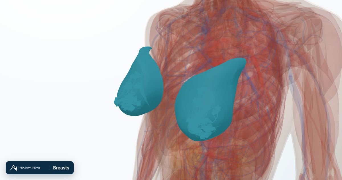
Breast cancer is the commonest cancer in women worldwide, and most cases occur after 50. Most tumours start in the milk ducts, fewer in the milk-producing lobules; ductal carcinoma in situ stays within the duct, while invasive cancer has broken through it. Every invasive cancer is tested for oestrogen receptors, progesterone receptors and the HER2 protein, which with grade and stage decides treatment. It is rare in men, and anyone with breast tissue can develop it.
Causes
- Accumulated DNA damage in duct or lobule cells, driven in part by lifetime exposure to oestrogen and progesterone
- Inherited faults in BRCA1, BRCA2 and a few other genes, responsible for a minority of cases
- Previous radiotherapy to the chest, particularly in youth
- No single cause is found in most cases; ageing is the strongest factor
Risk factors
- Increasing age: most cases are diagnosed after 50
- A close relative with breast or ovarian cancer, or a known BRCA1 or BRCA2 mutation
- Longer lifetime oestrogen exposure: an early first period, a late menopause, no pregnancies or a first pregnancy late in life
- Combined hormone replacement therapy and, to a small degree, the combined contraceptive pill: the increase is small, relates to how long they are used, and falls after stopping
- Alcohol, and being overweight after the menopause
- Dense breast tissue on mammography, a previous breast cancer or some benign breast conditions
- In trans women, several years of feminising hormone therapy: the risk is thought to be higher than in cis men but lower than in cis women; in men, conditions that raise oestrogen levels such as obesity, Klinefelter syndrome and cirrhosis
Symptoms
What a clinician may find
- A firm, irregular, usually painless lump in the breast or armpit
- Dimpling, puckering or an orange-peel texture of the skin over the breast
- A newly inverted nipple, a rash or crusting on the nipple, or blood-stained discharge
- A change in the size or outline of one breast
- In advanced disease, bone pain, breathlessness or jaundice from spread to bone, lung or liver
Complications
- Spread to the axillary lymph nodes and to bone, lung, liver or brain (metastatic breast cancer)
- Lymphoedema of the arm after axillary surgery or radiotherapy
- Bone loss and menopausal symptoms from endocrine therapy; heart effects of some chemotherapy and HER2-targeted drugs
- Recurrence in the breast, chest wall or elsewhere, sometimes many years later
- Anxiety, altered body image and loss of fertility in younger patients
Diagnosis
A suspicious lump or nipple change is assessed in a one-stop breast clinic by triple assessment: clinical examination, imaging (mammography, with ultrasound for younger women and dense breasts) and a core needle biopsy of anything abnormal. The biopsy confirms invasive or in situ disease, grade and receptor status (oestrogen receptor, progesterone receptor and HER2). Ultrasound of the axilla with needle sampling of suspicious nodes, and MRI in selected cases, complete local staging; CT and bone scans are reserved for larger tumours, involved nodes or symptoms suggesting spread. Genetic testing for BRCA1 and BRCA2 is offered when the age at diagnosis, tumour type or family history suggests an inherited cause. Screening in England invites women, and anyone else registered female with a GP who has breast tissue, for mammography every three years; the first invitation arrives between the ages of 50 and 53, routine invitations stop after 70, and anyone aged 71 or over can still ask their local breast screening service for screening. Trans women who have taken hormones for some years and trans men who have not had chest surgery may need to ask to be included.
Treatment
Early breast cancer is treated with surgery: either wide local excision, removing the tumour with a margin of normal tissue and followed by radiotherapy to the rest of the breast, or mastectomy, in each case with sentinel lymph node biopsy to check the axilla and clearance of the nodes if they are involved. Reconstruction can be immediate or delayed. Treatment given after surgery, or before it to shrink a large tumour, is chosen by receptor status and the risk of recurrence: chemotherapy; endocrine therapy for oestrogen-receptor-positive cancers (tamoxifen, or an aromatase inhibitor after the menopause), taken for years; and trastuzumab or similar antibodies for HER2-positive disease. Metastatic breast cancer is not usually curable but is often controlled for years with endocrine, targeted and chemotherapy, radiotherapy for painful bone deposits and bone-protecting drugs. Care is planned by a multidisciplinary team following NICE guidance.
Prevention
Not all breast cancer can be prevented, but keeping to a healthy weight, being physically active, limiting alcohol and breastfeeding all lower the risk, and hormone replacement therapy is best reviewed regularly and continued only for as long as it is needed. Attending screening finds cancers before they can be felt. People at high risk because of family history or a BRCA mutation can be offered earlier and more frequent imaging, risk-reducing medicines or risk-reducing surgery, planned with a genetics service.