



Imaging study · Moderate dose
CT scan
Also known as: computed tomography, CAT scan, CT angiography, CTPA, CT colonography
A rotating X-ray tube and computer build cross-sectional slices of the body in seconds, showing bone, organs, blood vessels and bleeding in fine detail; the workhorse of emergency imaging.
How it works
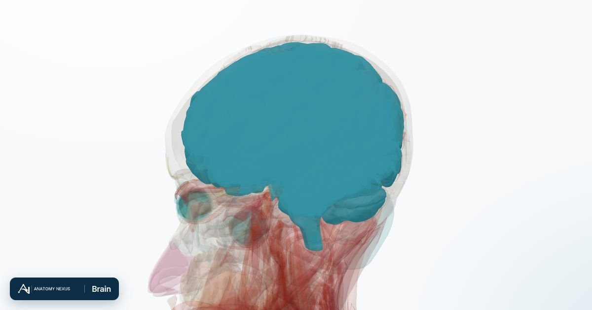
An X-ray tube spins around the body while it moves through the ring, collecting thousands of projections that a computer reconstructs into thin cross-sectional slices. The slices are stacked to view any plane or a 3D model. Iodine contrast injected into a vein lights up blood vessels and shows how organs and tumours take up blood.
What it shows
- Brain: bleeding, stroke (once established), tumours, injury
- Chest: lung nodules and cancer, blood clots in the lung arteries (CTPA), fibrosis, infection
- Abdomen and pelvis: appendicitis, diverticulitis, kidney stones, tumours, abscesses, bowel obstruction
- Bones: complex fractures, the spine and pelvis
- Blood vessels: aneurysms, dissection, coronary artery narrowing (CT coronary angiography)
Best for
- Emergencies where speed matters: trauma, stroke, suspected bleeding, severe abdominal pain
- Lung disease and cancer staging
- Kidney stones (no contrast needed)
- Detailed bone anatomy for surgical planning
Not the right tool for
- Early stroke (may look normal for hours), spinal cord, ligaments and cartilage (MRI)
- Repeated scans in children and young adults where dose accumulates
- People with severe kidney disease or contrast allergy (contrast may be withheld)
What to expect
- Modality
- CT (computed tomography)
- Preparation
- Fasting for a few hours before contrast; kidney function checked; a cannula placed. Oral contrast is sometimes drunk an hour before abdominal scans.
- Duration
- The scan itself takes seconds to a few minutes; the visit 20-30 minutes.
- Radiation
- A head CT is about 2 mSv, a chest CT 5-7 mSv and an abdomen-pelvis CT around 10 mSv: several years of background radiation, though modern scanners use far less than older ones.
- Contrast
- Iodine-based, injected into a vein for vessels, organs and tumours; causes a warm flush and a metallic taste. Metformin may be paused around the scan.
- Risks
- A small increase in lifetime cancer risk with each scan, weighed against the benefit; contrast reactions (rare) and a temporary drop in kidney function in those already at risk. Avoided in pregnancy unless essential.
Common uses
| Region | Typical question |
|---|---|
| Head | Head injury, sudden severe headache, first seizure, suspected stroke or bleed |
| Chest | Lung cancer staging, pulmonary embolism (CTPA), fibrosis, complicated pneumonia |
| Abdomen | Severe pain, suspected appendicitis, pancreatitis, bowel obstruction, cancer staging |
| Heart | CT coronary angiography for stable chest pain; calcium scoring |
Studies this page covers
CT headCT chestCT abdomenCT pelvisCT angiographyCT pulmonary angiographyCT colonographyLow-dose lung CTCT urogramCT perfusionHigh-resolution chest CTMusculoskeletal CT
Named studies of the master test taxonomy that this modality page covers; each links to its test category.