



Coronary Arteries Anatomy
Also known as: coronary arteries, coronary veins
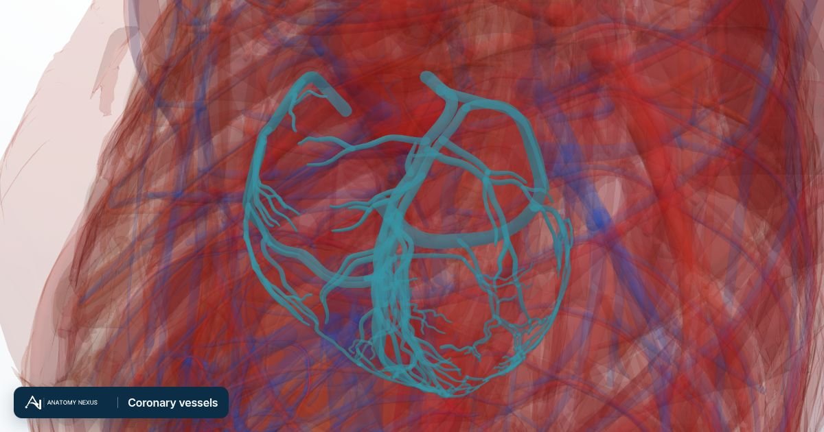
The coronary arteries are the heart's own blood supply: two vessels a few millimetres wide that arise from the aorta just above the aortic valve and run over the surface of the heart, sending branches into the muscle. Because each branch supplies a fixed territory, a blockage produces a predictable pattern of damage that the ECG, echocardiogram and angiogram can map.
Key facts
| Origin | Right and left aortic sinuses, just above the aortic valve |
|---|---|
| Left coronary artery | Short main stem dividing into the left anterior descending (anterior interventricular) and circumflex arteries |
| Right coronary artery | Runs in the right atrioventricular groove; gives the posterior interventricular artery in about 85% of people (right dominance) |
| Filling | Mainly during diastole, when the relaxed muscle lets blood through |
| Venous return | Great, middle and small cardiac veins to the coronary sinus |
| Flow | About 250 mL/min at rest, five times more in exercise |
Left coronary artery
The left main stem is only 1-2 cm long before it divides. The anterior interventricular branch, known clinically as the left anterior descending (LAD) artery, runs down the front of the interventricular groove to the apex, giving diagonal branches to the front of the left ventricle and septal branches into the septum; it supplies about half the left ventricle and most of the conduction bundles, which is why an LAD occlusion is the most dangerous heart attack. The circumflex artery curves round the left side in the atrioventricular groove, giving obtuse marginal branches to the lateral wall.
Right coronary artery
The right coronary artery runs in the right atrioventricular groove, giving a branch to the sinoatrial node in most people, acute marginal branches to the right ventricle, and, in about 85% of people, the posterior interventricular (posterior descending) artery and a branch to the atrioventricular node. It supplies the right ventricle, the inferior wall of the left ventricle and the conduction nodes, so its blockage causes inferior infarcts often complicated by slow rhythms.
Dominance and variation
The artery that gives off the posterior interventricular branch is called dominant: right in most people, left in about 8%, and shared in the rest. Anatomy varies enough that every angiogram is read individually. Collateral vessels between territories enlarge slowly when an artery narrows, which is why a chronic blockage can be survived while a sudden one is not.
Venous drainage
The great cardiac vein accompanies the anterior interventricular artery and curves round with the circumflex into the coronary sinus, which runs in the posterior atrioventricular groove and empties into the right atrium; the middle cardiac vein follows the posterior interventricular artery. Pacing leads for heart failure are threaded through the coronary sinus to reach the left ventricle.
Physiology
The coronary arteries are compressed by the contracting muscle, so most flow occurs in diastole. A fast heart rate shortens diastole and reduces supply just when demand rises. The vessels dilate in response to adenosine and nitric oxide released by working muscle; nitrates such as glyceryl trinitrate use the same pathway.
Clinical relevance
Fatty plaques in the wall narrow the lumen and cause angina on exertion; a ruptured plaque triggers a clot that blocks the artery and kills the muscle downstream within hours, a myocardial infarction. Troponin in the blood confirms damage, the ECG localises it, and an angiogram through a wrist or groin artery shows the blockage so that it can be opened with a balloon and stent or bypassed with a graft. Statins, aspirin and blood-pressure control slow the disease.
Structures in the atlas 26
The modelled pieces that make up the coronary vessels in the 3D atlas. Each opens in the explorer.
- Anterior cardiac vein · 2 pieces
- Anterior interventricular vein · 9 pieces
- Circumflex branch of left coronary artery · 6 pieces
- Conus branch of anterior interventricular branch of left coronary artery · 2 pieces
- Coronary sinus
- Diagonal branch of anterior descending branch of left coronary artery · 10 pieces
- First anterior ventricular branch of right coronary artery · 3 pieces
- First posterior ventricular branch of right coronary artery · 9 pieces
- First right anterior branch of anterior interventricular branch of left coronary artery · 2 pieces
- First septal branch of right posterior interventricular artery
- Great cardiac vein
- Left marginal vein · 3 pieces
- Marginal branch of right coronary artery · 5 pieces
- Middle cardiac vein · 14 pieces
- Posterior interventricular branch of right coronary artery · 9 pieces
- Posterior vein of left ventricle · 10 pieces
- Right conus artery · 2 pieces
- Right marginal vein · 3 pieces
- Second right anterior branch of anterior interventricular branch of left coronary artery
- Second septal branch of right posterior interventricular artery
- Septal branch of anterior interventricular artery · 3 pieces
- Small cardiac vein · 2 pieces
- Third right anterior branch of anterior interventricular branch of left coronary artery · 2 pieces
- Trunk of anterior interventricular branch of left coronary artery
- Trunk of left coronary artery
- Trunk of right coronary artery
The arteries in brief
Arteries carry blood away from the heart under pressure. The aorta leaves the left ventricle, arches over the heart giving off the vessels to the head and arms, then descends through the thorax and abdomen before dividing into the iliac arteries for the pelvis and legs. The pulmonary arteries carry deoxygenated blood from the right ventricle to the lungs. With 642 modelled pieces, this is the largest layer in the atlas, down to the segmental arteries of the lungs, liver and kidneys and the small branches on the surface of the brain.