



Aorta Anatomy
Also known as: aortic
The aorta is the largest artery in the body, a thick elastic tube about 2.5-3 cm wide that carries every drop of oxygenated blood from the left ventricle. It rises from the heart, arches over it, and descends through the chest and abdomen, giving branches to every organ before dividing into the two common iliac arteries.
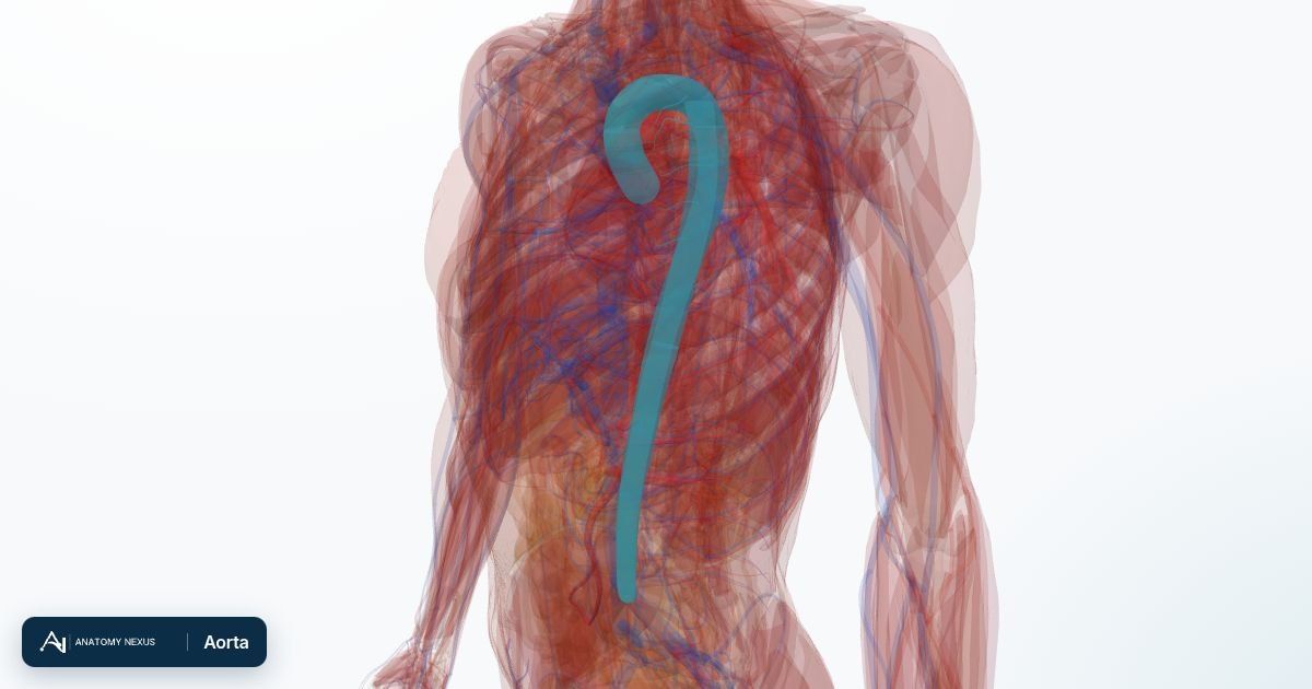
Key facts
| Segments | Ascending aorta, arch, descending thoracic aorta, abdominal aorta |
|---|---|
| Length | About 30-40 cm; ends at L4 by dividing into the common iliac arteries |
| Arch branches | Brachiocephalic trunk, left common carotid, left subclavian |
| Abdominal branches | Coeliac trunk, superior mesenteric, renal, gonadal and inferior mesenteric arteries |
| Wall | Three layers rich in elastin that store energy in systole and keep blood flowing in diastole |
| Pressure | About 120/80 mmHg at rest |
Ascending aorta
The aorta begins at the aortic valve inside the pericardium. Its first centimetres bulge into three aortic sinuses, from two of which the right and left coronary arteries arise, so the heart muscle is the first tissue the aorta supplies. The ascending aorta runs up and to the right for about 5 cm before curving into the arch.
Arch
The arch passes backwards and to the left over the left main bronchus and the pulmonary trunk, at the level of the sternal angle, giving off three great branches from its upper surface: the brachiocephalic trunk (which divides into the right common carotid and right subclavian arteries), the left common carotid artery and the left subclavian artery. Baroreceptors in the arch report blood pressure to the brainstem; the left recurrent laryngeal nerve hooks under it, so an enlarged arch can cause hoarseness.
Descending thoracic aorta
From the arch the aorta descends on the left side of the spine, then behind the oesophagus, to pass through the diaphragm at T12. It gives posterior intercostal arteries to the chest wall, bronchial arteries to the lungs, oesophageal branches and small vessels to the pericardium and diaphragm.
Abdominal aorta
In the abdomen the aorta lies to the left of the inferior vena cava, in front of the lumbar spine. Three unpaired branches supply the gut: the coeliac trunk (stomach, liver, spleen, pancreas), the superior mesenteric artery (small intestine and right colon) and the inferior mesenteric artery (left colon and rectum). Paired branches go to the adrenals, kidneys and gonads, and lumbar arteries to the back wall. At L4, level with the top of the hip bones, it divides into the common iliac arteries, which supply the pelvis and legs.
Wall structure and function
The aortic wall has an inner intima, a thick media of elastic lamellae and smooth muscle, and an outer adventitia carrying its own tiny vessels. During systole the elastic wall stretches to accept the ejected blood; during diastole it recoils and keeps blood flowing forward, smoothing the pulse. With age the elastin fragments, the aorta stiffens and systolic pressure rises.
Clinical relevance
Atherosclerosis roughens the aorta and its branches; cholesterol and blood pressure drive it. An aneurysm, most often of the abdominal aorta below the kidneys, can rupture with catastrophic bleeding, which is why men over 65 are offered a screening ultrasound. Aortic dissection, a tear in the intima that lets blood split the wall, causes tearing chest pain radiating to the back and is a surgical emergency. Coarctation is a congenital narrowing near the arch. Aortic valve stenosis obstructs the outlet from the heart.
Structures in the atlas 7
The modelled pieces that make up the aorta in the 3D atlas. Each opens in the explorer.
- Abdominal aorta
- Arch of aorta
- Ascending aorta
- Bronchial branch of arch of aorta
- Descending aorta
- Descending thoracic aorta
- Set of oesophageal branches of thoracic aorta
The arteries in brief
Arteries carry blood away from the heart under pressure. The aorta leaves the left ventricle, arches over the heart giving off the vessels to the head and arms, then descends through the thorax and abdomen before dividing into the iliac arteries for the pelvis and legs. The pulmonary arteries carry deoxygenated blood from the right ventricle to the lungs. With 642 modelled pieces, this is the largest layer in the atlas, down to the segmental arteries of the lungs, liver and kidneys and the small branches on the surface of the brain.