



Liver Anatomy
Also known as: hepatic
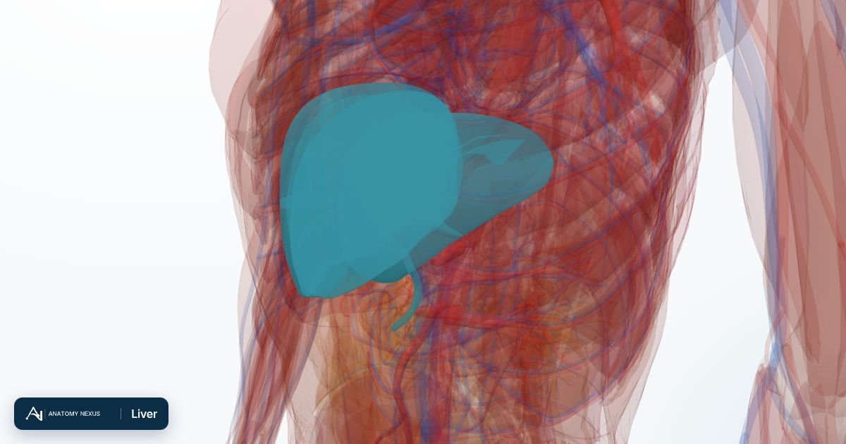
The liver is the largest internal organ, about 1.5 kg of chemical factory tucked under the right ribs. It receives nutrient-rich blood from the gut through the portal vein, processes and stores what the body needs, neutralises toxins, makes bile and most of the proteins in the blood, and can regenerate after losing more than half its mass.
Key facts
| Location | Right upper abdomen under the diaphragm, protected by ribs 7-11 |
|---|---|
| Weight | About 1.4-1.6 kg |
| Lobes | Right and left (with the small caudate and quadrate lobes); eight functional Couinaud segments |
| Blood supply | Hepatic portal vein (75%) and hepatic artery (25%), about 1.5 L/min |
| Drainage | Hepatic veins to the inferior vena cava; bile through the hepatic ducts |
| Cells | Hepatocytes arranged in plates around sinusoids; Kupffer cells filter the blood |
Location and surfaces
The liver lies against the underside of the diaphragm, mostly behind the right lower ribs, its left lobe reaching across to the stomach. Its smooth diaphragmatic surface is covered by peritoneum except for a bare area against the diaphragm; its visceral surface is moulded by the gallbladder, duodenum, right kidney and colon. The falciform ligament attaches it to the front abdominal wall and marks the boundary between the anatomical right and left lobes.
Lobes and segments
For surgery the liver is divided into eight Couinaud segments, each with its own branch of the portal vein, hepatic artery and bile duct, separated by planes that contain the hepatic veins. Segments II-IV form the functional left liver and V-VIII the right; segment I is the caudate lobe, which drains straight into the vena cava. A surgeon can remove any segment or group of segments along these planes with little bleeding and no loss of function in the remainder.
The porta hepatis and bile ducts
The portal vein, hepatic artery and common hepatic duct enter and leave the liver at the porta hepatis on its underside, wrapped together in the hepatoduodenal ligament. Bile made by hepatocytes flows through tiny canaliculi into ductules and then the right and left hepatic ducts, which join as the common hepatic duct; the cystic duct from the gallbladder joins it to form the common bile duct, which opens into the duodenum beside the pancreatic duct.
Blood supply
Uniquely, most of the liver's blood is venous: the hepatic portal vein brings blood that has already passed through the stomach, intestines, pancreas and spleen, loaded with absorbed nutrients, gut hormones and anything else taken by mouth, including drugs and toxins. The hepatic artery, a branch of the coeliac trunk, brings oxygenated blood. Both mix in the sinusoids between plates of hepatocytes. Because everything absorbed from the gut passes through the liver first, oral drugs can be partly destroyed before reaching the circulation (the first-pass effect).
Venous drainage
Blood leaves through three hepatic veins that open into the inferior vena cava just below the diaphragm. When the liver is scarred by cirrhosis, resistance rises, pressure backs up in the portal system, and blood finds alternative routes through veins around the oesophagus and stomach, which can bleed catastrophically.
Innervation
Sympathetic fibres from the coeliac plexus and parasympathetic fibres from the vagus reach the liver along its vessels. The liver's own tissue is insensitive to pain; the ache of a swollen liver comes from stretch of its capsule and the overlying peritoneum, and is often felt at the right shoulder because the phrenic nerve shares spinal segments with the shoulder skin.
Function
The liver stores glucose as glycogen and releases it between meals, makes new glucose from amino acids, builds plasma proteins including albumin and the clotting factors, manufactures cholesterol and bile salts, stores iron and vitamins A, D and B12, breaks down worn-out red cells into bilirubin, converts ammonia to urea, and inactivates hormones, alcohol and most drugs through its cytochrome enzymes.
Clinical relevance
Fatty liver, alcohol and viral hepatitis inflame and eventually scar the liver; cirrhosis blocks portal flow and cripples synthesis, so patients bleed, swell with ascites and become jaundiced and confused. Liver function tests read these processes in the blood. Gallstones can block the common bile duct, causing jaundice and pancreatitis. The liver is the commonest site for cancer to spread through the portal vein from the bowel. Paracetamol overdose destroys hepatocytes within days.
Structures in the atlas 22
The modelled pieces that make up the liver in the 3D atlas. Each opens in the explorer.
- Anterior inferior tributary of right hepatic biliary tree
- Anterior superior tributary of right hepatic biliary tree
- Caudate lobe of liver
- Caudate lobe tributary of left hepatic biliary tree
- Common hepatic duct
- Hepatovenous segment II
- Hepatovenous segment III
- Hepatovenous segment IV
- Hepatovenous segment IX
- Hepatovenous segment V
- Hepatovenous segment VI
- Hepatovenous segment VII
- Hepatovenous segment VIII · 2 pieces
- Lateral inferior tributary of left hepatic biliary tree
- Lateral superior tributary of left hepatic biliary tree · 2 pieces
- Left duct of caudate lobe of liver
- Left hepatic duct
- Medial inferior tributary of left hepatic biliary tree
- Medial superior tributary of left hepatic biliary tree · 2 pieces
- Posterior inferior tributary of right hepatic biliary tree
- Posterior superior tributary of right hepatic biliary tree · 2 pieces
- Right hepatic duct
The digestive in brief
The digestive tract is a nine-metre tube from mouth to anus. Food is chewed, swallowed down the oesophagus, churned with acid in the stomach, digested and absorbed along the small intestine (duodenum, jejunum, ileum), and its residue concentrated in the colon. The liver, gallbladder and pancreas pour bile and enzymes into the duodenum. The model includes the tongue, salivary glands and gums, the stomach and intestines in many segments, the liver as eight hepatovenous segments with its bile ducts, the gallbladder, the pancreas and the mesenteries that suspend the gut.