



Stomach Anatomy
Also known as: gastric
The stomach is a muscular, J-shaped bag in the upper left abdomen that receives food from the oesophagus, churns it with acid and enzymes into a paste called chyme, and releases it in small amounts into the duodenum. Its lining secretes about two litres of gastric juice a day and protects itself with a layer of mucus and bicarbonate.
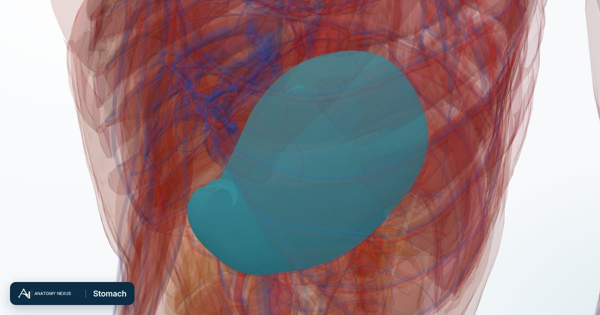
Key facts
| Location | Left upper abdomen under the diaphragm, between the oesophagus and duodenum |
|---|---|
| Regions | Cardia, fundus, body, antrum, pylorus |
| Capacity | About 50 mL empty, 1-1.5 L after a meal, up to 4 L |
| Secretions | Hydrochloric acid, pepsinogen, intrinsic factor, mucus, gastrin, ghrelin |
| Blood supply | Branches of the coeliac trunk: left and right gastric, gastro-epiploic and short gastric arteries |
| Nerve supply | Vagus nerves (secretion, motility) and sympathetic fibres from the coeliac plexus |
Location and relations
The stomach lies in the left hypochondrium and epigastrium, below the diaphragm and the left lobe of the liver, in front of the pancreas, spleen and left kidney. Its lesser curvature faces the liver and its greater curvature carries the apron-like greater omentum. The lower oesophageal sphincter at the cardia stops acid refluxing upward; the pyloric sphincter at the outlet controls emptying into the duodenum.
Regions
The cardia surrounds the oesophageal opening; the fundus is the dome that rises above it, often full of swallowed air; the body is the main reservoir; the antrum grinds food against the closed pylorus and releases the hormone gastrin; and the pylorus is the thick muscular outlet. Ulcers favour the lesser curvature and the antrum; the fundus and body carry the acid-secreting glands.
Wall and lining
Three layers of smooth muscle (an extra oblique layer inside the usual circular and longitudinal coats) let the stomach churn as well as squeeze. The mucosa is thrown into folds (rugae) that flatten as it fills and is packed with gastric pits leading to glands: parietal cells make hydrochloric acid and intrinsic factor (needed to absorb vitamin B12), chief cells make pepsinogen, and mucous cells coat the surface with alkaline mucus. Proton pump inhibitors shut down the acid pump in the parietal cells.
Blood supply
All of the stomach's arteries come from the coeliac trunk: the left gastric runs along the lesser curvature to meet the right gastric from the hepatic artery, the right and left gastro-epiploic arteries run along the greater curvature, and short gastric arteries from the splenic artery reach the fundus. This rich supply is why bleeding ulcers can be life-threatening, and why the stomach can be reshaped in surgery without losing its blood supply.
Venous drainage
Gastric veins accompany the arteries and drain into the portal vein, so stomach blood reaches the liver first. In cirrhosis, veins around the cardia enlarge into varices that can rupture.
Innervation
The anterior and posterior vagal trunks enter the abdomen on the oesophagus and supply the stomach: they trigger acid secretion in anticipation of food and drive the peristaltic waves that mix and empty it. Sympathetic fibres reduce motility and carry pain, which is felt in the epigastrium.
Function
The stomach stores a meal, kills most swallowed microbes with acid at pH 1-2, begins protein digestion with pepsin, mixes everything into chyme and meters it into the duodenum at a rate the small intestine can handle, roughly 2-4 hours for a mixed meal. It signals hunger with ghrelin and fullness through stretch receptors.
Clinical relevance
Acid reflux irritates the oesophagus when the lower sphincter fails; Helicobacter pylori infection and anti-inflammatory drugs breach the mucus barrier and cause gastritis and peptic ulcers; stomach cancer arises from the lining. A gastroscopy passes a camera through the cardia to inspect every region and take biopsies.
About
The J-shaped muscular sac in the upper left abdomen where food is stored and churned with acid and pepsin.
Function. Begins protein digestion, kills microbes with acid and releases chyme into the duodenum in measured amounts.
Clinical note. Peptic ulcers (often from Helicobacter pylori), gastritis and gastric cancer.
Structures in the atlas 1
The modelled pieces that make up the stomach in the 3D atlas. Each opens in the explorer.
The digestive in brief
The digestive tract is a nine-metre tube from mouth to anus. Food is chewed, swallowed down the oesophagus, churned with acid in the stomach, digested and absorbed along the small intestine (duodenum, jejunum, ileum), and its residue concentrated in the colon. The liver, gallbladder and pancreas pour bile and enzymes into the duodenum. The model includes the tongue, salivary glands and gums, the stomach and intestines in many segments, the liver as eight hepatovenous segments with its bile ducts, the gallbladder, the pancreas and the mesenteries that suspend the gut.