



Pancreas Anatomy
Also known as: pancreatic
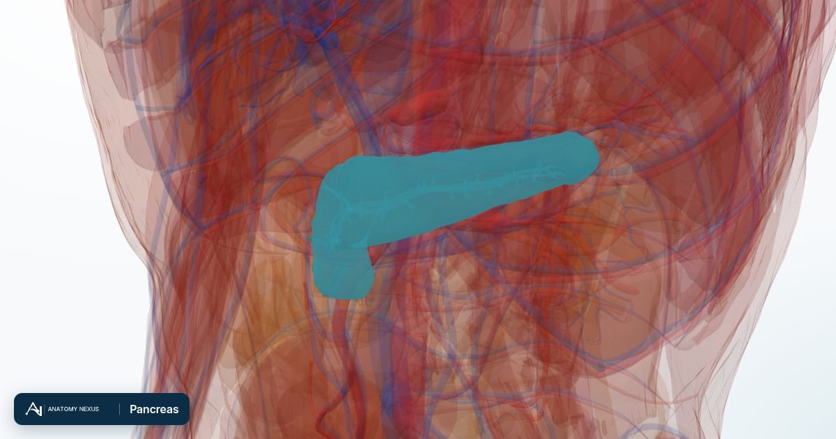
The pancreas is a soft, elongated gland about 15 cm long lying across the back of the upper abdomen behind the stomach. It is two organs in one: exocrine tissue that pours digestive enzymes into the duodenum, and scattered islets that release insulin and glucagon into the blood to control glucose.
Key facts
| Location | Retroperitoneal, across L1-L2, head in the curve of the duodenum, tail reaching the spleen |
|---|---|
| Parts | Head (with uncinate process), neck, body, tail |
| Ducts | Main pancreatic duct joining the common bile duct at the ampulla; accessory duct |
| Exocrine output | About 1.5 L of alkaline enzyme-rich juice a day |
| Endocrine cells | About a million islets of Langerhans: beta cells (insulin), alpha cells (glucagon), delta cells (somatostatin) |
| Blood supply | Pancreaticoduodenal arteries and branches of the splenic artery |
Location and relations
The pancreas lies behind the peritoneum of the lesser sac, with the stomach in front of it and the aorta, inferior vena cava, left kidney and spine behind. Its head nestles in the C-shaped curve of the duodenum, the neck lies over the superior mesenteric vessels, and the tail reaches the hilum of the spleen. Being so deep, the pancreas is hard to examine and its disease often presents late; its pain radiates through to the back.
Ducts
The main pancreatic duct runs the length of the gland collecting from lobular ducts and, in most people, joins the common bile duct to open into the duodenum at the major papilla, guarded by the sphincter of Oddi. A gallstone lodging here blocks both ducts, causing jaundice and pancreatitis. The smaller accessory duct opens separately a little higher.
Exocrine tissue
Acinar cells make inactive enzyme precursors (trypsinogen, chymotrypsinogen, lipase, amylase) and duct cells add bicarbonate-rich fluid that neutralises stomach acid. The enzymes are only activated in the duodenum, a safeguard that fails in pancreatitis, when the gland digests itself.
Islets
Islets of Langerhans, more numerous in the tail, sense blood glucose directly. Beta cells release insulin after meals so that muscle, fat and liver take up glucose; alpha cells release glucagon between meals to mobilise it. Autoimmune destruction of beta cells causes type 1 diabetes; resistance to insulin with failing beta-cell reserve causes type 2.
Blood supply
The head is supplied by the superior pancreaticoduodenal arteries from the gastroduodenal artery and the inferior pancreaticoduodenal arteries from the superior mesenteric artery, which form arcades shared with the duodenum, so the two cannot be separated in surgery (the Whipple operation removes both). The body and tail receive branches from the splenic artery as it runs along the upper border. Veins drain to the splenic and superior mesenteric veins and thus the portal vein.
Innervation
Vagal fibres stimulate secretion; sympathetic fibres from the coeliac and superior mesenteric plexuses carry pain to T6-T10, which is why pancreatic pain bores through to the mid-back. Coeliac plexus block is used to relieve pain in pancreatic cancer.
Clinical relevance
Acute pancreatitis, usually from gallstones or alcohol, causes severe upper abdominal pain and a raised blood lipase or amylase. Chronic pancreatitis destroys both functions, causing diabetes and fatty stools. Pancreatic cancer in the head presents with painless jaundice from bile duct obstruction. Blood glucose and HbA1c measure the islets' work.
Structures in the atlas 4
The modelled pieces that make up the pancreas in the 3D atlas. Each opens in the explorer.
The digestive in brief
The digestive tract is a nine-metre tube from mouth to anus. Food is chewed, swallowed down the oesophagus, churned with acid in the stomach, digested and absorbed along the small intestine (duodenum, jejunum, ileum), and its residue concentrated in the colon. The liver, gallbladder and pancreas pour bile and enzymes into the duodenum. The model includes the tongue, salivary glands and gums, the stomach and intestines in many segments, the liver as eight hepatovenous segments with its bile ducts, the gallbladder, the pancreas and the mesenteries that suspend the gut.