



Imaging study · No radiation
MRI scan
Also known as: magnetic resonance imaging, MR scan, MR angiography, functional MRI, cardiac MRI
Magnetic resonance imaging uses a strong magnet and radio waves, not radiation, to produce detailed pictures of soft tissue: the brain, spinal cord, joints, muscles and organs.
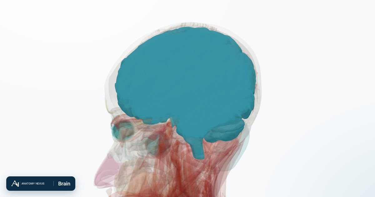
How it works
A powerful magnet aligns the hydrogen nuclei in the body's water and fat. Radio pulses knock them out of alignment and, as they relax back, they emit faint signals that differ by tissue type. Different pulse sequences (T1, T2, diffusion, fat suppression) emphasise different properties, giving unrivalled soft-tissue contrast without ionising radiation.
What it shows
- Brain and spinal cord: stroke within minutes (diffusion), multiple sclerosis, tumours, epilepsy foci, dementia patterns
- Spine: disc herniation, nerve compression, infection, cord injury
- Joints: cartilage, menisci, ligaments, tendons, bone marrow oedema (early stress fractures)
- Heart: muscle scar, cardiomyopathy, congenital disease
- Liver, pancreas, bile ducts (MRCP), prostate, pelvis, breast
Best for
- Any question about the brain, spinal cord or nerves
- Ligament, cartilage and tendon injuries of the knee, shoulder and ankle
- Characterising liver, pancreatic and prostate lesions
- Children and pregnancy, where radiation is avoided
Not the right tool for
- Emergencies needing speed (a scan takes 20-60 minutes and the environment is hard to monitor)
- Lungs and calcified structures (few hydrogen nuclei; CT is better)
- People with certain implants, severe claustrophobia, or who cannot lie still
What to expect
- Modality
- MRI (magnetic resonance imaging)
- Preparation
- A detailed safety questionnaire about implants, metal fragments, pacemakers and previous surgery; all metal removed; ear protection for the loud knocking. Fasting for some abdominal scans.
- Duration
- 20-60 minutes lying still inside a tube; each sequence lasts a few minutes.
- Radiation
- No ionising radiation; the magnetic field and radio waves have no known lasting effect at diagnostic strengths.
- Contrast
- Gadolinium, injected into a vein, highlights tumours, inflammation and blood vessels; rare allergic reactions.
- Risks
- Ferromagnetic objects become projectiles; older pacemakers, cochlear implants, aneurysm clips and metal in the eye can be dangerous. Gadolinium contrast is avoided in severe kidney disease and pregnancy. Claustrophobia is the commonest problem.
Common uses
| Region | Typical question |
|---|---|
| Brain | Stroke, tumours, multiple sclerosis, epilepsy, dementia, headache with red flags |
| Spine | Sciatica with weakness, suspected cauda equina, infection, myelopathy |
| Knee and shoulder | Ligament and meniscal tears, rotator cuff tears, cartilage |
| Abdomen and pelvis | Liver lesions, bile duct stones (MRCP), prostate cancer, endometriosis, rectal cancer staging |
Studies this page covers
Brain MRISpine MRICardiac MRIBreast MRIAbdominal MRIPelvic MRIMusculoskeletal MRIMR angiographyMR venographyMagnetic resonance cholangiopancreatographyFunctional MRIDiffusion MRIPerfusion MRIMR spectroscopyProstate MRI
Named studies of the master test taxonomy that this modality page covers; each links to its test category.