



Spine Anatomy
Also known as: spine, backbone, vertebrae, vertebral column, spinal column
The vertebral column is a stack of 33 vertebrae, 24 of them movable, separated by shock-absorbing discs and bound by ligaments and muscles. It carries the weight of the head and trunk, allows the trunk to bend and twist, and shelters the spinal cord and the nerve roots that leave it at every level.
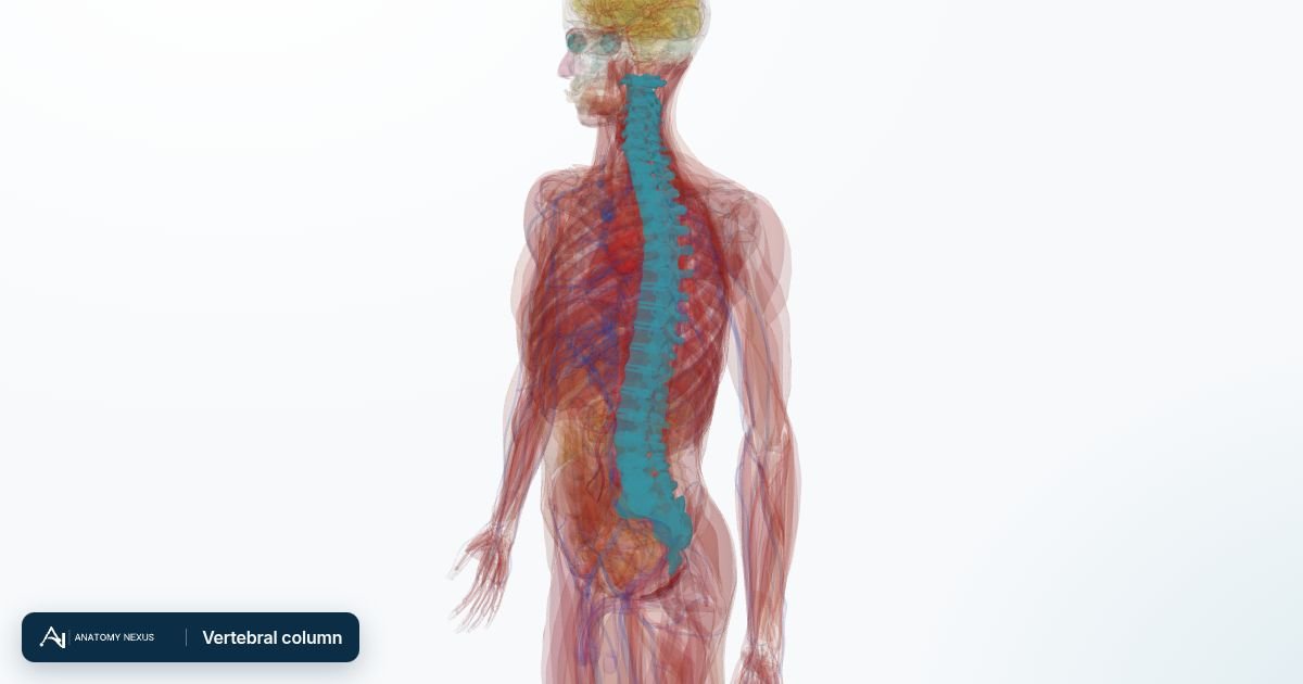
Key facts
| Vertebrae | 7 cervical, 12 thoracic, 5 lumbar, 5 fused sacral, 4 fused coccygeal |
|---|---|
| Curves | Cervical and lumbar lordosis, thoracic and sacral kyphosis |
| Discs | 23 intervertebral discs, about a quarter of the column's height |
| Spinal cord | Ends at about L1-L2 in adults; nerve roots continue as the cauda equina |
| Nerves | 31 pairs of spinal nerves leaving through the intervertebral foramina |
| Movement | Most flexion and extension in the cervical and lumbar spine; rotation mainly thoracic |
A typical vertebra
Each movable vertebra has a weight-bearing body in front and a bony arch behind that encloses the vertebral foramen; stacked, these foramina form the vertebral canal for the spinal cord. The arch carries a spinous process (the bump felt under the skin), two transverse processes and four articular facets that form small synovial joints with the vertebrae above and below. The facet joints guide movement and can wear like any other joint.
Regions
The cervical vertebrae are small, with holes in their transverse processes for the vertebral arteries; the atlas (C1) supports the skull and nods on it, the axis (C2) provides the peg on which the head turns. Thoracic vertebrae have facets for the ribs and long sloping spines, so the thoracic spine is stiff. Lumbar vertebrae are massive to carry the body's weight and allow bending. The sacrum wedges between the hip bones to transmit weight to the legs, and the coccyx is the vestigial tail.
Intervertebral discs
Between the bodies sit discs made of a tough ring of fibrocartilage (annulus fibrosus) around a gel-like nucleus pulposus. They absorb shock, spread load and permit small movements that add up to considerable flexibility. With age the nucleus dries and the annulus cracks; a herniated disc pushes nucleus material backwards into a nerve root, most often at L4-L5 or L5-S1, causing sciatica.
Ligaments and muscles
The anterior and posterior longitudinal ligaments run the length of the bodies; the ligamentum flavum joins the arches; supraspinous and interspinous ligaments link the spines. Deep muscles (erector spinae, multifidus) extend and stabilise the spine; the abdominal muscles and psoas flex it. Most back pain arises from these muscles, ligaments and facet joints rather than the discs.
Spinal cord and nerves
The spinal cord runs from the medulla to about the first lumbar vertebra, wrapped in the same three meninges as the brain and bathed in cerebrospinal fluid, which is why a lumbar puncture below L2 can sample the fluid safely. Thirty-one pairs of spinal nerves leave through the intervertebral foramina; each carries motor fibres to a group of muscles and sensation from a strip of skin (a dermatome). Below the cord the roots hang as the cauda equina; compression there is a surgical emergency.
Blood supply
Segmental arteries from the vertebral, intercostal and lumbar arteries supply the vertebrae and send radicular branches to the cord, which also has a single anterior and two posterior spinal arteries along its length. One large radicular artery in the lower thoracic region (the artery of Adamkiewicz) supplies most of the lower cord, and its injury during aortic surgery can cause paralysis.
Clinical relevance
Mechanical back pain, disc herniation, spinal stenosis, osteoporotic vertebral fractures and spondylolisthesis are the common spinal problems. Red flags such as leg weakness, numbness in the saddle area or loss of bladder control point to cord or cauda equina compression and need an urgent MRI. Injuries are assessed by the level of the vertebra and the spinal segment affected.
Structures in the atlas 48
The modelled pieces that make up the vertebral column in the 3D atlas. Each opens in the explorer.
- Atlas
- Axis
- Eighth thoracic vertebra
- Eleventh thoracic vertebra
- Fifth cervical vertebra
- Fifth lumbar vertebra
- Fifth thoracic vertebra
- First lumbar vertebra
- First thoracic vertebra
- Fourth cervical vertebra
- Fourth lumbar vertebra
- Fourth thoracic vertebra
- Intervertebral disk
- Intervertebral disk of axis
- Intervertebral disk of eighth thoracic vertebra
- Intervertebral disk of eleventh thoracic vertebra
- Intervertebral disk of fifth cervical vertebra
- Intervertebral disk of fifth lumbar vertebra
- Intervertebral disk of fifth thoracic vertebra
- Intervertebral disk of first lumbar vertebra
- Intervertebral disk of first thoracic vertebra
- Intervertebral disk of fourth cervical vertebra
- Intervertebral disk of fourth lumbar vertebra
- Intervertebral disk of fourth thoracic vertebra
- Intervertebral disk of ninth thoracic vertebra
- Intervertebral disk of second lumbar vertebra
- Intervertebral disk of second thoracic vertebra
- Intervertebral disk of seventh cervical vertebra
- Intervertebral disk of seventh thoracic vertebra
- Intervertebral disk of sixth cervical vertebra
- Intervertebral disk of sixth thoracic vertebra
- Intervertebral disk of tenth thoracic vertebra
- Intervertebral disk of third cervical vertebra
- Intervertebral disk of third lumbar vertebra
- Intervertebral disk of third thoracic vertebra
- Ninth thoracic vertebra
- Sacrum
- Second lumbar vertebra
- Second thoracic vertebra
- Seventh cervical vertebra
- Seventh thoracic vertebra
- Sixth cervical vertebra
- Sixth thoracic vertebra
- Tenth thoracic vertebra
- Third cervical vertebra
- Third lumbar vertebra
- Third thoracic vertebra
- Twelfth thoracic vertebra
The skeleton in brief
The adult skeleton has 206 bones plus the cartilages that cap joints, join the ribs to the sternum and shape the nose and airway. It is organised into an axial skeleton (skull, vertebral column, ribs and sternum) that protects the brain, spinal cord, heart and lungs, and an appendicular skeleton (the limbs with their girdles) built for movement. Bone is living tissue: it remodels throughout life in response to load, stores calcium and phosphate, and houses the marrow that makes blood cells.