



Imaging study · Low dose
DXA scan (bone density)
Also known as: DEXA scan, bone density scan, bone densitometry, dual-energy X-ray absorptiometry
A quick, very low-dose X-ray measurement of bone mineral density at the hip and spine, reported as a T-score, that diagnoses osteoporosis and estimates fracture risk.
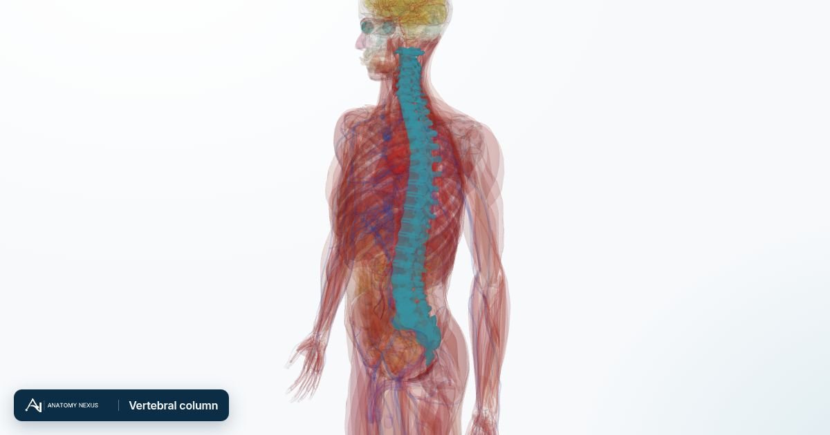
How it works
Two X-ray beams of different energies pass through the bone; the difference in how much each is absorbed gives the amount of mineral per area of bone. The scanner passes over the lower spine and hip while the person lies still on a padded table for about 10-20 minutes.
What it shows
- Bone mineral density at the hip and lumbar spine, and sometimes the forearm
- A T-score comparing the result with young healthy adults and a Z-score comparing it with people of the same age and sex
- Change in density on repeat scans, to monitor treatment
Best for
- Diagnosing osteoporosis and osteopenia
- Estimating fracture risk together with clinical risk factors (FRAX)
- Deciding on and monitoring bone-protective treatment
- Assessing bone loss from long-term corticosteroids or other causes
Not the right tool for
- Finding the cause of bone or back pain
- Detecting fractures, tumours or arthritis in detail: X-ray, CT or MRI do that
- Children and people with spinal hardware, where interpretation needs specialist adjustment
What to expect
- Modality
- DXA bone densitometry
- Preparation
- No calcium supplements on the day of the scan; wear clothes without metal; mention recent contrast X-rays or nuclear scans, which can interfere.
- Duration
- 10-20 minutes
- Radiation
- Very low: a fraction of a chest X-ray, less than a day of natural background radiation.
- Contrast
- None
- Risks
- Minimal radiation exposure; not done in pregnancy unless essential.
Common uses
| Region | Typical question |
|---|---|
| Hip and spine | Bone density for osteoporosis diagnosis and fracture risk |
| Forearm | When hip or spine cannot be measured, or in hyperparathyroidism |
| Whole body | Body composition (fat and lean mass) in specialist settings |
Studies this page covers
Named studies of the master test taxonomy that this modality page covers; each links to its test category.