



Knee Joint Anatomy
Also known as: knee, knee joint, knees
The knee is the largest joint in the body: a hinge between the femur and tibia, with the patella gliding in front, held together by four main ligaments and cushioned by two menisci. It bears several times body weight with every step and is the joint most often injured in sport and most often replaced in later life.
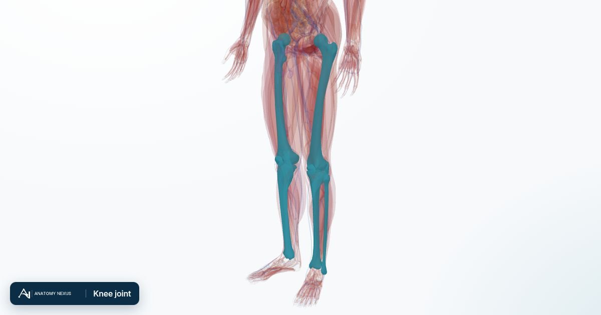
Key facts
| Type | Modified hinge (synovial) joint: flexion, extension and a little rotation |
|---|---|
| Bones | Femoral condyles, tibial plateau, patella; the fibula lies alongside but is not part of the joint |
| Ligaments | Anterior and posterior cruciate, medial and lateral collateral, patellar ligament |
| Menisci | Medial (C-shaped) and lateral (more circular) fibrocartilage pads |
| Movement | About 0-140 degrees of flexion; the joint locks in full extension |
| Blood supply | Genicular branches of the popliteal artery |
Bones and surfaces
The rounded femoral condyles roll and glide on the almost flat tibial plateau, an unstable arrangement that depends entirely on ligaments and muscles for stability. The patella, a sesamoid bone in the quadriceps tendon, slides in the groove between the condyles and increases the leverage of the quadriceps. The head of the fibula articulates with the tibia below the joint line and anchors the lateral collateral ligament.
Ligaments
Inside the joint the anterior cruciate ligament (ACL) stops the tibia sliding forward and limits rotation; the posterior cruciate stops it sliding back. The medial collateral ligament resists the knee being forced inwards (valgus) and blends with the medial meniscus; the lateral collateral is a separate cord resisting varus force. The patellar ligament carries the quadriceps pull from the patella to the tibial tuberosity. A twisting fall tears the ACL with a pop and rapid swelling; a blow to the outside of the knee sprains the medial collateral.
Menisci
Two crescents of fibrocartilage deepen the tibial surface, spread the load over the cartilage and absorb shock. Their outer third has a blood supply and can heal; the inner part does not. Tears cause pain along the joint line, catching and locking, and are seen on MRI or arthroscopy.
Capsule, bursae and synovium
A fibrous capsule lined with synovial membrane surrounds the joint and produces the lubricating fluid; an effusion (water on the knee) stretches it. Several bursae reduce friction, including the prepatellar bursa in front of the kneecap (housemaid's knee when inflamed) and a bursa behind the knee that can swell into a Baker's cyst.
Muscles and movement
The quadriceps extend the knee and the hamstrings flex it; the gastrocnemius, popliteus and the iliotibial tract also cross the joint. In the last degrees of extension the femur rotates medially on the tibia, screwing the joint tight so that standing needs little muscular effort; the popliteus unlocks it. Strong quadriceps and hamstrings are the best protection for the ligaments.
Blood and nerve supply
The popliteal artery passes behind the knee and sends genicular branches around the joint that form an anastomosis, a safety net if the main artery is compressed. The tibial and common fibular nerves pass behind and beside the joint; the common fibular nerve winds round the fibular neck under the skin, where it is easily bruised, causing foot drop. Joint pain is carried by branches of the femoral, obturator, tibial and common fibular nerves, so hip disease can refer pain to the knee.
Clinical relevance
Osteoarthritis wears away the cartilage, most often in the medial compartment, causing pain, stiffness and bowing; X-rays show joint-space narrowing and a knee replacement resurfaces the worn ends. Ligament and meniscal injuries are diagnosed by examination and MRI. A hot swollen knee may be gout, pseudogout or infection, and a needle aspiration of the fluid tells them apart.
Structures in the atlas 10
The modelled pieces that make up the knee joint in the 3D atlas. Each opens in the explorer.
- Left femur
- Left fibula
- Left patella
- Left popliteus
- Left tibia
- Right femur
- Right fibula
- Right patella
- Right popliteus
- Right tibia
The skeleton in brief
The adult skeleton has 206 bones plus the cartilages that cap joints, join the ribs to the sternum and shape the nose and airway. It is organised into an axial skeleton (skull, vertebral column, ribs and sternum) that protects the brain, spinal cord, heart and lungs, and an appendicular skeleton (the limbs with their girdles) built for movement. Bone is living tissue: it remodels throughout life in response to load, stores calcium and phosphate, and houses the marrow that makes blood cells.