



Brain Anatomy
Also known as: cerebrum
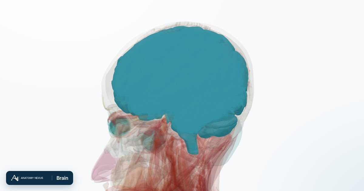
The brain is about 1.4 kg of nervous tissue floating in cerebrospinal fluid inside the skull. Its wrinkled cerebral hemispheres handle perception, movement, language, memory and planning; the cerebellum coordinates movement; and the brainstem keeps breathing, heart rate and consciousness going.
Key facts
| Weight | About 1.3-1.4 kg, 2% of body weight, using 20% of the body's oxygen |
|---|---|
| Main parts | Cerebrum (two hemispheres), diencephalon (thalamus, hypothalamus), brainstem (midbrain, pons, medulla), cerebellum |
| Lobes | Frontal, parietal, temporal, occipital, plus the insula and limbic lobe |
| Blood supply | Two internal carotid and two vertebral arteries joining at the circle of Willis |
| Protection | Skull, three meninges and about 150 mL of cerebrospinal fluid |
| Neurons | Roughly 86 billion |
Location and coverings
The brain fills the cranial cavity and is wrapped in three meninges: the tough dura mater lining the skull, the spidery arachnoid, and the delicate pia mater clinging to every fold. Cerebrospinal fluid made in the ventricles circulates in the subarachnoid space between arachnoid and pia, cushioning the brain and reducing its effective weight to a few tens of grams. The brain leaves the skull through the foramen magnum as the spinal cord.
Cerebral hemispheres and lobes
Each hemisphere is folded into gyri separated by sulci, which triples the surface area of the cortex. The central sulcus divides the frontal lobe (movement, planning, speech production in Broca's area, personality) from the parietal lobe (touch, spatial awareness); the lateral sulcus marks off the temporal lobe (hearing, language comprehension in Wernicke's area, memory via the hippocampus); the occipital lobe at the back is vision. The precentral gyrus is the primary motor cortex and the postcentral gyrus the primary somatosensory cortex, each mapping the opposite side of the body. The corpus callosum, a thick band of white matter, connects the two hemispheres.
Deep structures
Beneath the cortex lie the basal ganglia (caudate nucleus, putamen, globus pallidus), which shape movement and are lost in Parkinson's disease; the thalamus, the relay station for almost all sensation on its way to the cortex; and the hypothalamus, which controls hunger, thirst, temperature, sleep and the pituitary gland below it. The hippocampus and amygdala in the medial temporal lobe form new memories and process emotion.
Brainstem and cerebellum
The midbrain, pons and medulla oblongata carry every tract between the brain and spinal cord and house the nuclei of ten of the twelve cranial nerves, the breathing and cardiovascular centres, and the reticular formation that maintains wakefulness. The cerebellum, tucked beneath the occipital lobes, compares intended with actual movement and corrects it, giving smooth, coordinated action and balance.
Blood supply
The internal carotid arteries supply the front two-thirds of each hemisphere through the anterior and middle cerebral arteries; the vertebral arteries join to form the basilar artery, which supplies the brainstem, cerebellum and, through the posterior cerebral arteries, the occipital lobes. These vessels meet at the circle of Willis on the underside of the brain, a ring that can partly compensate for a blocked artery. The middle cerebral artery is the vessel most often blocked in stroke; because it supplies the motor and language areas, its blockage causes the face, arm and speech signs of the FAST test.
Venous drainage
Cerebral veins drain into the dural venous sinuses, channels between the layers of dura: the superior sagittal sinus along the top of the brain, the transverse and sigmoid sinuses curving down to become the internal jugular veins. Cerebrospinal fluid is absorbed into the same sinuses through arachnoid granulations.
Function
Sensory information from the body crosses to the opposite hemisphere, so the left brain controls and feels the right side of the body. Language is usually left-hemisphere dominant. Neurons communicate with electrical impulses and chemical neurotransmitters; the pattern of connections, refined throughout life, underlies learning and memory.
Clinical relevance
Stroke, tumours, trauma, infection and degenerative diseases such as Alzheimer's and Parkinson's each damage particular regions, and the resulting deficits map back onto the anatomy: weakness from the motor cortex or internal capsule, memory loss from the hippocampus, tremor and rigidity from the basal ganglia, unsteadiness from the cerebellum. Raised pressure inside the rigid skull, from bleeding or swelling, is dangerous because the brain can be pushed through the openings in the dura and the foramen magnum.
Structures in the atlas 95
The modelled pieces that make up the brain in the 3D atlas. Each opens in the explorer.
- Anterior commissure
- Anterior part of left superior temporal gyrus
- Anterior part of right superior temporal gyrus
- Brachium of left inferior colliculus
- Brachium of left superior colliculus
- Brachium of right inferior colliculus
- Brachium of right superior colliculus
- Cerebellum · 2 pieces
- Cerebral aqueduct
- Choroid plexus of cerebral hemisphere · 2 pieces
- Commissure of fornix of forebrain
- Corpus callosum
- Fourth ventricle
- Habenula
- Hypothalamus · 2 pieces
- Interpeduncular fossa · 2 pieces
- Interventricular foramen
- Lamina terminalis · 2 pieces
- Left amygdala
- Left angular gyrus
- Left caudate nucleus
- Left cingulate gyrus
- Left fornix of forebrain
- Left fusiform gyrus
- Left globus pallidus
- Left hippocampus
- Left inferior colliculus
- Left inferior frontal gyrus
- Left inferior temporal gyrus
- Left insula
- Left internal capsule
- Left lateral geniculate body
- Left lateral ventricle
- Left medial geniculate body
- Left middle frontal gyrus
- Left middle temporal gyrus
- Left occipital lobe
- Left optic tract
- Left parahippocampal gyrus
- Left postcentral gyrus
- Left precentral gyrus
- Left putamen
- Left stria medullaris of thalamus
- Left stria terminalis
- Left superior colliculus
- Left superior frontal gyrus
- Left supramarginal gyrus
- Left thalamus
- Mammillary body · 2 pieces
- Medulla oblongata · 2 pieces
- Midbrain · 2 pieces
- Optic chiasm · 2 pieces
- Orbital gyrus · 2 pieces
- Peduncle of midbrain · 2 pieces
- Pineal body
- Pons · 2 pieces
- Posterior commissure
- Posterior part of left superior temporal gyrus
- Posterior part of right superior temporal gyrus
- Right amygdala
- Right angular gyrus
- Right caudate nucleus
- Right cingulate gyrus
- Right fornix of forebrain
- Right fusiform gyrus
- Right globus pallidus
- Right hippocampus
- Right inferior colliculus
- Right inferior frontal gyrus
- Right inferior temporal gyrus
- Right insula
- Right internal capsule
- Right lateral geniculate body
- Right lateral ventricle
- Right medial geniculate body
- Right middle frontal gyrus
- Right middle temporal gyrus
- Right occipital lobe
- Right optic tract
- Right parahippocampal gyrus
- Right postcentral gyrus
- Right precentral gyrus
- Right putamen
- Right stria medullaris of thalamus
- Right superior colliculus
- Right superior frontal gyrus
- Right supramarginal gyrus
- Right thalamus
- Septum of telencephalon
- Stria terminalis
- Tentorium cerebelli
- Third ventricle
- Tuber cinereum · 2 pieces
- White matter of left cerebral hemisphere
- White matter of right cerebral hemisphere
The nervous system in brief
The nervous system gathers information, decides and commands. The brain and spinal cord form the central nervous system; the cranial and spinal nerves with their ganglia form the peripheral nervous system. This model is richest in the head: the cerebral gyri and lobes, the basal ganglia, thalamus and hypothalamus, the brainstem and cerebellum, the ventricles, the optic pathway and the nerves of the orbit. Peripheral nerves of the limbs are not part of this BodyParts3D release.