



Heart Anatomy
Also known as: cardiac
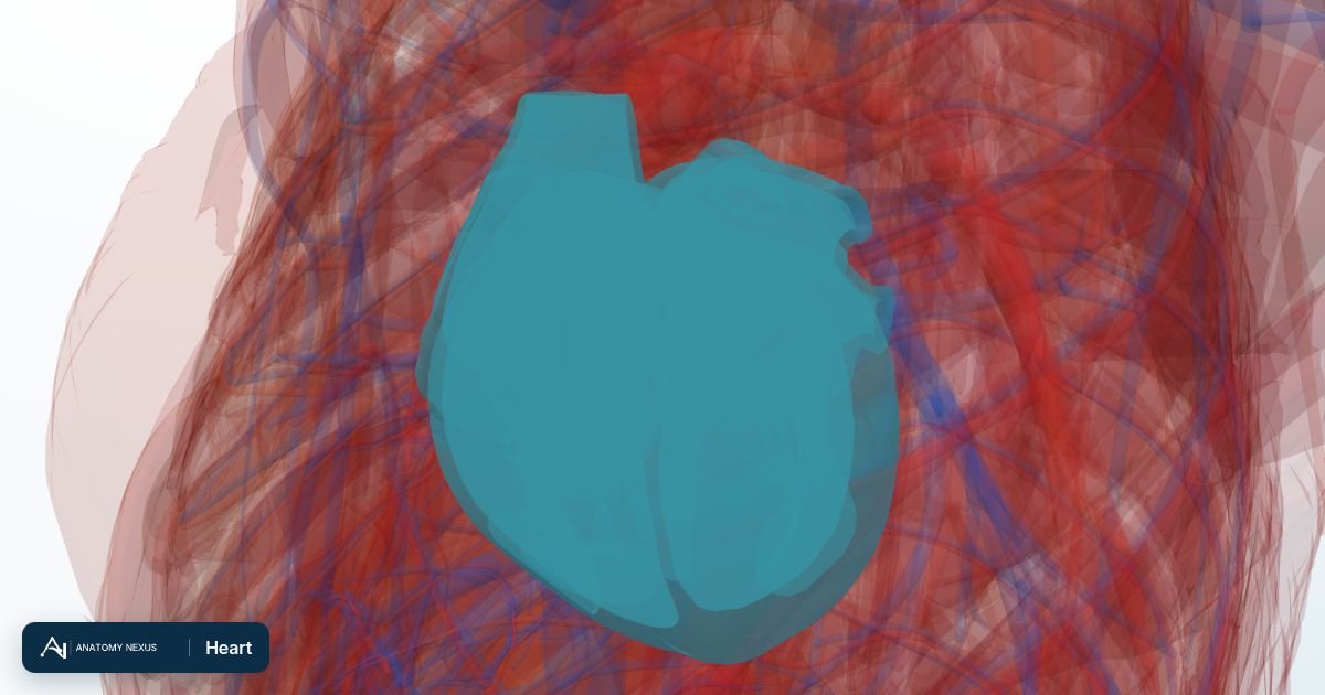
The heart is a fist-sized muscular pump in the middle of the chest, tilted so that two-thirds of it lies to the left of the midline. Its four chambers and four valves move about five litres of blood every minute at rest, sending deoxygenated blood to the lungs and oxygenated blood to the rest of the body.
Key facts
| Location | Middle mediastinum, behind the sternum, between the lungs, resting on the diaphragm |
|---|---|
| Chambers | Right atrium, right ventricle, left atrium, left ventricle |
| Valves | Tricuspid, pulmonary, mitral, aortic |
| Weight | About 250-350 g in adults |
| Blood supply | Right and left coronary arteries from the ascending aorta |
| Nerve supply | Sympathetic fibres (speed up) and the vagus nerve (slow down) via the cardiac plexus |
| Output | About 5 L/min at rest, up to 25 L/min in exercise |
Location and coverings
The heart sits in the middle mediastinum, enclosed in the pericardium: a tough fibrous outer sac fused to the diaphragm below and the great vessels above, lined by a double layer of serous membrane whose thin film of fluid lets the heart beat almost without friction. The apex points down and to the left, reaching the fifth intercostal space in the midclavicular line, where the apex beat can be felt. The base faces backwards, towards the oesophagus and descending aorta, which is why an oesophageal ultrasound probe gives such clear views of the atria.
Chambers
The right atrium receives deoxygenated blood from the superior and inferior venae cavae and the coronary sinus, and passes it through the tricuspid valve into the right ventricle, a thin-walled crescent-shaped chamber that pumps into the pulmonary trunk. The left atrium receives oxygenated blood through four pulmonary veins and fills the left ventricle, whose wall is about three times thicker than the right because it must generate pressures of 120 mmHg or more to drive blood around the whole body. The interatrial and interventricular septa separate the two sides; the foramen ovale, open before birth, leaves a small depression in the interatrial septum.
Valves
Two atrioventricular valves (the tricuspid on the right and the mitral, with two leaflets, on the left) are held closed during ventricular contraction by chordae tendineae anchored to papillary muscles, so they cannot balloon back into the atria. Two semilunar valves (pulmonary and aortic), each with three cusps, prevent blood ejected from the ventricles from flowing back. The valve rings sit in a fibrous skeleton that also insulates the atria electrically from the ventricles, so that the only electrical route between them is the atrioventricular node and bundle.
Blood supply
The right and left coronary arteries arise from the aortic sinuses just above the aortic valve and fill mainly during diastole. The left coronary artery divides within a few centimetres into the anterior interventricular (left anterior descending) artery, running down the front of the interventricular septum, and the circumflex artery, curving round in the left atrioventricular groove. The right coronary artery runs in the right atrioventricular groove, usually giving off the sinoatrial nodal artery and, in most people, the posterior interventricular artery that supplies the inferior wall. Blockage of any of these branches starves a predictable territory of muscle.
Venous drainage
Most venous blood from the heart wall collects in the coronary sinus, a wide vein in the posterior atrioventricular groove that empties into the right atrium; the great, middle and small cardiac veins are its main tributaries. Small anterior cardiac veins and the tiny thebesian veins drain directly into the chambers.
Innervation and conduction
The heart beats on its own: the sinoatrial node in the right atrial wall near the superior vena cava fires about 70 times a minute, the impulse spreads across the atria to the atrioventricular node, pauses, then races down the bundle of His and its branches to the ventricular muscle. The autonomic nervous system only modulates this rhythm. Sympathetic fibres from the upper thoracic spinal cord increase rate and force; parasympathetic fibres in the vagus nerve slow it. Pain from the heart is referred to the chest, left arm, neck and jaw because those areas share spinal cord segments with the cardiac sympathetic nerves.
Function
Each cardiac cycle has a filling phase (diastole) and a contraction phase (systole). The right side of the heart sends blood through the pulmonary circulation to pick up oxygen and unload carbon dioxide; the left side sends it through the systemic circulation. Cardiac output is heart rate multiplied by stroke volume, and both are adjusted continuously by nerves, hormones and the volume of blood returning to the heart.
Clinical relevance
Narrowing of the coronary arteries causes angina and heart attacks; the territory affected shows on the ECG and echocardiogram. Valve disease (a leaking mitral valve, a narrowed aortic valve) can be heard as murmurs and measured with ultrasound. Failure of the pump causes fluid to back up into the lungs and legs. Disorders of the conduction system cause slow, fast or irregular rhythms such as atrial fibrillation. Cardiopulmonary resuscitation works because the heart lies directly behind the lower sternum, where compressions squeeze it against the spine.
Structures in the atlas 20
The modelled pieces that make up the heart in the 3D atlas. Each opens in the explorer.
- Anterior leaflet of mitral valve
- Anterior leaflet of tricuspid valve
- Anterior papillary muscle of right ventricle
- Anterolateral head of lateral papillary muscle of left ventricle
- Cavity of left atrium
- Cavity of left ventricle
- Cavity of right atrium
- Cavity of right ventricle
- Lateral papillary muscle of left ventricle
- Left anterior cusp of pulmonary valve
- Posterior cusp of pulmonary valve
- Posterior leaflet of mitral valve
- Posterior leaflet of tricuspid valve
- Posterior papillary muscle of right ventricle
- Right anterior cusp of pulmonary valve
- Septal leaflet of tricuspid valve
- Septal papillary muscle of right ventricle
- Wall of left atrium
- Wall of right atrium
- Wall of ventricle
The heart in brief
The heart is a four-chambered muscular pump about the size of a fist. The right atrium receives deoxygenated blood from the venae cavae and passes it through the tricuspid valve to the right ventricle, which pumps it to the lungs. Oxygenated blood returns to the left atrium, crosses the mitral valve into the thick-walled left ventricle and is ejected through the aortic valve into the aorta. The model separates the atrial and ventricular walls, the chambers, the valve leaflets and cusps, and the papillary muscles that anchor the mitral and tricuspid valves.