



Body system · 20 pieces
Heart
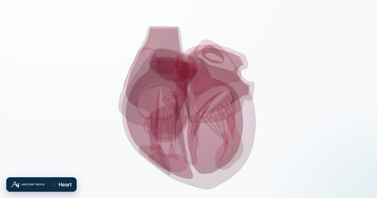
Chambers, valves and walls of the heart, with the pericardium.
Overview
The heart is a four-chambered muscular pump about the size of a fist. The right atrium receives deoxygenated blood from the venae cavae and passes it through the tricuspid valve to the right ventricle, which pumps it to the lungs. Oxygenated blood returns to the left atrium, crosses the mitral valve into the thick-walled left ventricle and is ejected through the aortic valve into the aorta. The model separates the atrial and ventricular walls, the chambers, the valve leaflets and cusps, and the papillary muscles that anchor the mitral and tricuspid valves.
Functions
- Pump blood through the pulmonary and systemic circulations
- Keep blood flowing one way with four valves
- Generate its own rhythm through the conduction system
Clinical notes
- Coronary artery disease and myocardial infarction
- Heart failure when the ventricles cannot pump enough
- Valve disease: stenosis or regurgitation
- Arrhythmias such as atrial fibrillation
Organs & groups in this system
All structures 20
20 modelled pieces. Click a structure to open it in the atlas.
- Anterior leaflet of mitral valve
- Anterior leaflet of tricuspid valve
- Anterior papillary muscle of right ventricle
- Anterolateral head of lateral papillary muscle of left ventricle
- Cavity of left atrium
- Cavity of left ventricle
- Cavity of right atrium
- Cavity of right ventricle
- Lateral papillary muscle of left ventricle
- Left anterior cusp of pulmonary valve
- Posterior cusp of pulmonary valve
- Posterior leaflet of mitral valve
- Posterior leaflet of tricuspid valve
- Posterior papillary muscle of right ventricle
- Right anterior cusp of pulmonary valve
- Septal leaflet of tricuspid valve
- Septal papillary muscle of right ventricle
- Wall of left atrium
- Wall of right atrium
- Wall of ventricle