



Large Intestine Anatomy
Also known as: colon, bowel, rectum, appendix, caecum
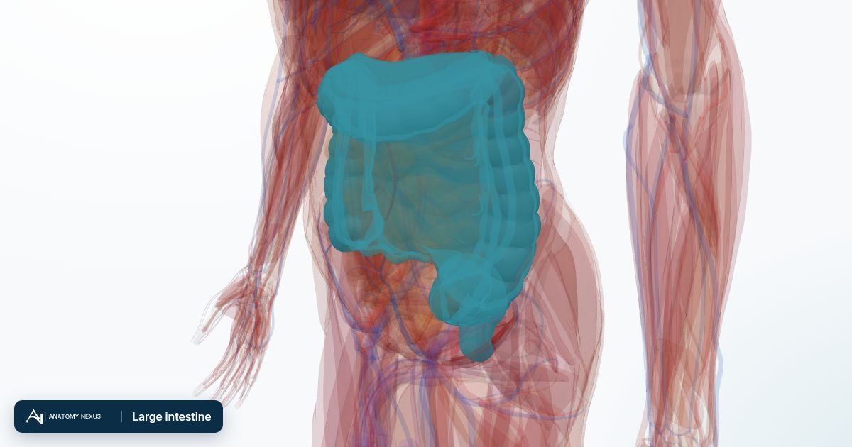
The large intestine is the last 1.5 metres of the gut: the caecum with its appendix, the ascending, transverse, descending and sigmoid colon, the rectum and the anal canal. It absorbs water and salts from the liquid residue arriving from the small intestine, hosts trillions of bacteria, and stores and expels the stool.
Key facts
| Length | About 1.5 m, 6-7 cm wide at the caecum narrowing to 2.5 cm at the sigmoid |
|---|---|
| Parts | Caecum and appendix, ascending, transverse, descending and sigmoid colon, rectum, anal canal |
| Blood supply | Superior mesenteric artery to the caecum, ascending and most of the transverse colon; inferior mesenteric artery to the rest |
| Nerve supply | Vagus as far as the transverse colon, pelvic parasympathetic nerves beyond; sympathetic fibres from the mesenteric plexuses |
| Transit time | About 12-48 hours |
| Water absorbed | About 1-1.5 L a day, leaving 100-200 g of stool |
Caecum and appendix
The ileum opens into the caecum in the right lower abdomen through the ileocaecal valve, which stops contents flowing backwards. The appendix, a blind tube 5-10 cm long, hangs from the caecum; its position varies, most often behind the caecum, which is why appendicitis can present in unusual ways. Its wall is rich in lymphoid tissue.
Colon
The ascending colon runs up the right side to the hepatic flexure under the liver, the transverse colon hangs across the abdomen on its own mesentery to the splenic flexure, and the descending colon runs down the left side to become the S-shaped sigmoid colon in the pelvis. The colon's outer muscle is gathered into three bands (taeniae coli) that pucker it into pouches (haustra) and carry fatty tags (appendices epiploicae). The lining has no villi; it is a smooth mucosa packed with mucus-secreting glands.
Rectum and anal canal
The rectum follows the curve of the sacrum, storing stool until defecation, and passes through the pelvic floor into the anal canal, closed by an involuntary internal sphincter of smooth muscle and a voluntary external sphincter of skeletal muscle. Stretch of the rectum triggers the urge to defecate; the sphincters and the puborectalis muscle allow it to be deferred.
Blood supply
The superior mesenteric artery supplies the caecum, ascending colon and two-thirds of the transverse colon through the ileocolic, right colic and middle colic arteries; the inferior mesenteric artery supplies the rest through the left colic, sigmoid and superior rectal arteries. The marginal artery runs along the whole colon linking the two systems. The splenic flexure, at the junction of the two supplies, is the part most vulnerable when blood flow falls.
Venous drainage
Veins mirror the arteries and drain into the portal vein, so colonic cancers spread first to the liver. The lower rectum and anal canal drain into systemic veins as well, so cancers there can spread to the lungs, and the connection between the two systems forms haemorrhoids when pressure rises.
Innervation
Parasympathetic fibres (vagus for the proximal colon, pelvic splanchnic nerves from S2-S4 for the descending colon and rectum) drive peristalsis and secretion; sympathetic fibres slow the gut and carry pain. Because pain from the gut is referred to the midline, appendicitis begins as vague pain around the navel and only moves to the right lower abdomen when the inflamed appendix touches the parietal peritoneum.
Function
The colon absorbs water and sodium, converting about 1.5 litres of liquid chyme into formed stool. Its bacteria ferment undigested fibre into short-chain fatty acids that feed the lining, make vitamin K and some B vitamins, and shape the immune system. Mass movements a few times a day propel contents into the rectum.
Clinical relevance
Bowel cancer, most often in the rectum and sigmoid, arises from polyps that a colonoscopy can remove; screening uses a stool test for hidden blood. Ulcerative colitis inflames the rectum and colon, Crohn's disease any part of the gut. Diverticula form in the sigmoid where pressure is highest. Appendicitis is the commonest surgical emergency.
Structures in the atlas 11
The modelled pieces that make up the large intestine in the 3D atlas. Each opens in the explorer.
- Appendix
- Ascending colon
- Descending colon
- Marginal artery of colon
- Mesoappendix
- Rectum
- Taenia libera
- Taenia mesocolica
- Taenia omentalis
- Transverse colon
- Transverse mesocolon
The digestive in brief
The digestive tract is a nine-metre tube from mouth to anus. Food is chewed, swallowed down the oesophagus, churned with acid in the stomach, digested and absorbed along the small intestine (duodenum, jejunum, ileum), and its residue concentrated in the colon. The liver, gallbladder and pancreas pour bile and enzymes into the duodenum. The model includes the tongue, salivary glands and gums, the stomach and intestines in many segments, the liver as eight hepatovenous segments with its bile ducts, the gallbladder, the pancreas and the mesenteries that suspend the gut.