



Procedure · Endoscopic procedures
Colonoscopy
Also known as: lower endoscopy, bowel scope, flexible sigmoidoscopy, bowel cancer screening colonoscopy
A flexible camera is passed through the anus around the whole large intestine to the caecum and often into the last part of the small intestine. Polyps found on the way are removed, which prevents them growing into cancer; this is why screening colonoscopy reduces bowel cancer deaths.
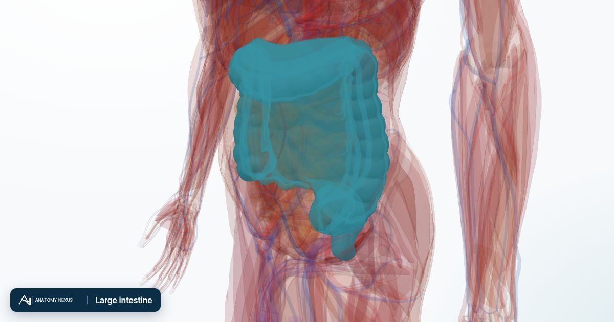
What it is
A flexible camera is passed through the anus around the whole large intestine to the caecum and often into the last part of the small intestine. Polyps found on the way are removed, which prevents them growing into cancer; this is why screening colonoscopy reduces bowel cancer deaths.
Why it is done
- A positive stool screening test (FIT) or bleeding from the bowel
- Change in bowel habit, unexplained weight loss or iron-deficiency anaemia
- Diagnosing and monitoring inflammatory bowel disease
- Surveillance after polyps or bowel cancer, and in high-risk families
- Removing polyps, stopping bleeding, dilating strictures, decompressing a twisted bowel
Before the procedure
- Low-fibre diet for a few days, then strong laxative bowel preparation the day before, drinking litres of clear fluid until the output runs clear
- Iron tablets stopped a week before; blood thinners and diabetes medicines adjusted
- Sedation and painkiller through a cannula, or gas and air
Step by step
- Lying on the left side; the lubricated scope is inserted and advanced with carbon dioxide inflating the bowel
- The scope is steered around the sigmoid, descending, transverse and ascending colon to the caecum, changing position or with abdominal pressure to straighten loops
- The lining is inspected carefully on the slow withdrawal (at least six minutes)
- Polyps are snared with a wire loop and cut with diathermy, or removed cold if small; larger lesions lifted and resected in pieces
- Biopsies are taken from any abnormal or inflamed areas
Afterwards
- Wind and cramps for a few hours as gas passes
- Eat and drink normally; a little blood after polypectomy is expected
- No driving for 24 hours after sedation
- Histology in one to two weeks decides surveillance intervals
Recovery
Normal activities the next day.
Risks
- Bleeding after polyp removal (about 1 in 100 for large polyps, sometimes delayed by a week)
- Perforation (about 1 in 1000-2000), more after resection of large polyps; may need surgery
- Missed lesions (some per cent, especially with poor preparation)
- Reaction to sedation; dehydration from bowel preparation in the frail
Alternatives
- CT colonography (virtual colonoscopy): no sedation, but no biopsies and needs colonoscopy if abnormal
- Flexible sigmoidoscopy: examines only the left colon with an enema
- Capsule colonoscopy in selected cases
Educational content. A general description of how a procedure is usually done. Details vary between hospitals, surgeons and patients; the treating team explains the specifics and the consent.