



Bronchoscopy
Also known as: airway endoscopy, EBUS, endobronchial ultrasound, flexible bronchoscopy
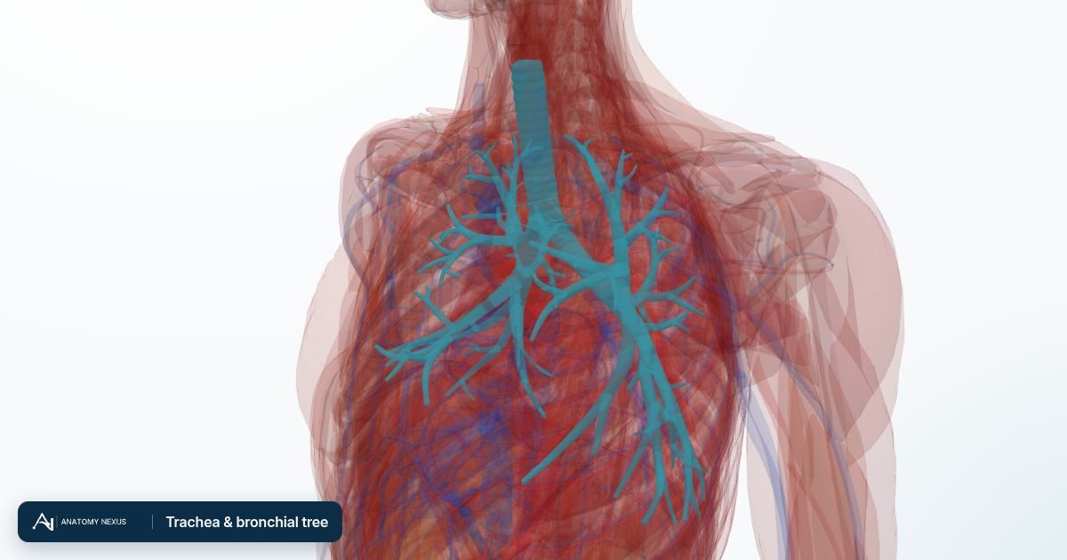
A thin flexible scope is passed through the nose or mouth, between the vocal cords and down the trachea into the bronchi of each lung, to inspect the airways, wash out or brush cells, and take biopsies. Endobronchial ultrasound (EBUS) adds an ultrasound probe on the scope tip so lymph nodes beside the airways can be sampled with a needle through the wall.
What it is
A thin flexible scope is passed through the nose or mouth, between the vocal cords and down the trachea into the bronchi of each lung, to inspect the airways, wash out or brush cells, and take biopsies. Endobronchial ultrasound (EBUS) adds an ultrasound probe on the scope tip so lymph nodes beside the airways can be sampled with a needle through the wall.
Why it is done
- A lung mass or shadow on CT, to diagnose and stage lung cancer
- Coughing up blood
- Persistent infection or collapse of a lung segment; sampling in immunosuppressed patients
- Removing inhaled foreign bodies and mucus plugs; placing airway stents
- Diagnosing sarcoidosis, tuberculosis and interstitial lung disease (cryobiopsy)
Before the procedure
- CT of the chest; clotting and blood count; spirometry and oxygen level
- Fasting 4-6 hours; blood thinners managed
- Local anaesthetic spray to the nose and throat plus sedation; oxygen through the nose; monitoring throughout
Step by step
- The scope passes through the nostril, over the back of the tongue, and lidocaine is sprayed onto the vocal cords before passing between them
- The trachea and the bronchial tree of both lungs are inspected to segmental level
- Saline washings (lavage) are instilled and suctioned for microbiology and cytology; brushings scrape cells from a lesion
- Forceps biopsies are taken from visible tumours; transbronchial biopsies from lung tissue beyond view, sometimes under X-ray guidance
- With EBUS, a needle passes through the airway wall into lymph nodes under ultrasound view
Afterwards
- Nothing to eat or drink until the throat numbness wears off (1-2 hours)
- Mild cough, sore throat, a little blood-streaked sputum for a day
- A chest X-ray if transbronchial biopsies were taken, to check for a collapsed lung
- No driving for 24 hours; results in a week or two
Recovery
Normal activities the next day.
Risks
- Low oxygen and breathlessness during the procedure
- Bleeding, usually minor
- Pneumothorax (collapsed lung) after transbronchial biopsy (a few per cent)
- Fever, infection, arrhythmia, laryngospasm (rare)
Alternatives
- CT-guided needle biopsy through the chest wall for peripheral lesions
- Sputum cytology and blood tests
- Surgical biopsy (mediastinoscopy, VATS)