



Lung Anatomy
Also known as: lung, pulmonary
The lungs are two spongy, cone-shaped organs filling most of the chest on either side of the heart. Air reaches them through a branching tree of airways ending in about 300 million alveoli, whose combined surface, roughly the size of a tennis court, is where oxygen enters the blood and carbon dioxide leaves it.
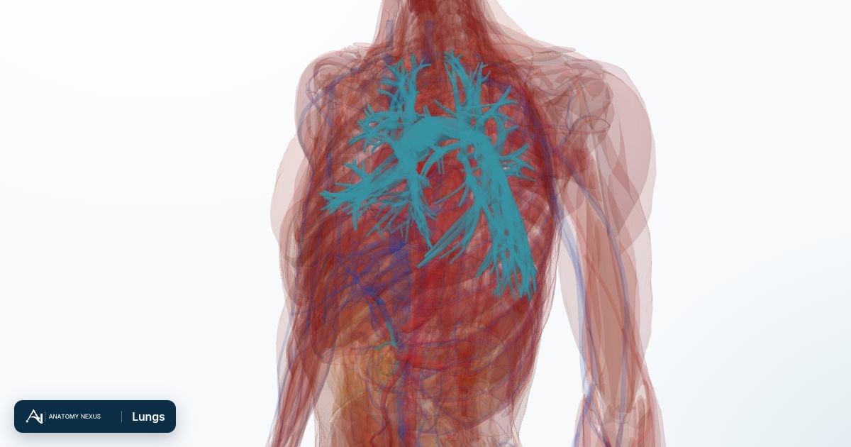
Key facts
| Lobes | Three on the right (upper, middle, lower), two on the left (upper, lower) with a lingula |
|---|---|
| Airways | Trachea, main bronchi, lobar and segmental bronchi, bronchioles, alveolar ducts, alveoli |
| Segments | 10 bronchopulmonary segments per lung, each with its own bronchus and artery |
| Blood supply | Pulmonary arteries (for gas exchange) and bronchial arteries (for the lung tissue itself) |
| Nerve supply | Vagus nerve (constricts airways, mucus) and sympathetic fibres (dilate airways) |
| Breathing | About 12-16 breaths a minute at rest, 500 mL each |
Location and pleura
Each lung sits in its own pleural cavity, wrapped in visceral pleura that is continuous with the parietal pleura lining the chest wall, diaphragm and mediastinum. The thin film of fluid between the two layers lets the lung slide as the chest expands and holds it against the chest wall by surface tension, so that when the diaphragm descends the lung is pulled open. Air entering this space (a pneumothorax) breaks the seal and the lung collapses. The right lung is larger and shorter, pushed up by the liver; the left has a cardiac notch that makes room for the heart.
Lobes and segments
Fissures divide the right lung into upper, middle and lower lobes and the left into upper and lower lobes. Each lobe is built from bronchopulmonary segments, pyramids of lung tissue with their own segmental bronchus and artery and no connection to their neighbours, which is why surgeons can remove one segment and radiologists describe a shadow by segment.
Airways
The trachea divides at the carina into right and left main bronchi. The right is wider and more vertical, so inhaled objects usually go that way. Each main bronchus divides into lobar, then segmental bronchi, then some twenty further generations of ever-smaller bronchi and bronchioles. Cartilage rings stiffen the larger airways; smooth muscle in the bronchioles adjusts their calibre, tightening in asthma. Ciliated cells and mucus sweep dust and microbes upward to be swallowed. Beyond the terminal bronchioles, respiratory bronchioles and alveolar ducts open into clusters of alveoli.
Alveoli and gas exchange
An alveolus is a thin-walled sac about 0.2 mm across, lined by flat type I cells for gas exchange and type II cells that secrete surfactant, the detergent that stops the sacs collapsing at the end of each breath. Capillaries form a dense net around every alveolus, so the barrier between air and blood is under a micrometre thick. Oxygen diffuses into red cells and binds haemoglobin; carbon dioxide diffuses the other way and is breathed out.
Blood supply
The pulmonary trunk from the right ventricle divides into right and left pulmonary arteries, which follow the bronchi into every segment and carry deoxygenated blood to the alveolar capillaries. Four pulmonary veins return oxygenated blood to the left atrium. Separately, small bronchial arteries from the aorta nourish the bronchi and lung tissue. A clot lodging in a pulmonary artery (pulmonary embolism) blocks flow to the segment it serves.
Innervation
The pulmonary plexus around each lung root carries parasympathetic fibres from the vagus nerve, which constrict the airways and stimulate mucus, and sympathetic fibres, which relax airway muscle. Salbutamol and adrenaline relieve wheeze by acting on the same beta-2 receptors that the sympathetic nerves use. The lung itself has no pain fibres; chest pain from lung disease comes from the parietal pleura, supplied by the intercostal and phrenic nerves.
Function
Breathing in is active: the diaphragm flattens and the external intercostals lift the ribs, enlarging the chest so air flows in. Breathing out at rest is passive elastic recoil. The brainstem sets the rhythm, driven mainly by the level of carbon dioxide in the blood. Beyond gas exchange the lungs filter small clots, make angiotensin-converting enzyme, and defend against inhaled microbes.
Clinical relevance
Asthma and COPD narrow the airways; pneumonia fills alveoli with fluid and pus; fibrosis stiffens the lung; emphysema destroys alveolar walls; cancer most often arises from the bronchial lining. Spirometry measures how much and how fast air moves; a chest X-ray and CT show the lobes, segments and pleural spaces; a pulse oximeter reads how well the alveoli are loading oxygen onto haemoglobin.
Structures in the atlas 81
The modelled pieces that make up the lungs in the 3D atlas. Each opens in the explorer.
- Accessory subsuperior branch of right posterior basal segmental artery · 2 pieces
- Anterior branch of lateral segmental artery · 2 pieces
- Anterior branch of right anterior segmental artery · 4 pieces
- Anterior part of lateral segmental vein · 3 pieces
- Anterior part of right apical segmental artery · 4 pieces
- Anterior part of right apical segmental vein · 2 pieces
- Apical part of right apical segmental artery · 4 pieces
- Apical part of right apical segmental vein · 3 pieces
- Apical part of right posterior segmental artery · 3 pieces
- Basal branch of right anterior basal segmental artery · 2 pieces
- Inferior branch of medial segmental artery · 2 pieces
- Inferior lingular artery · 2 pieces
- Inferior lingular bronchial tree · 2 pieces
- Inferior lingular vein · 2 pieces
- Inferior part of medial segmental vein · 2 pieces
- Inferior part of right anterior segmental vein
- Lateral branch of right anterior basal segmental artery
- Lateral branch of right lateral basal segmental artery · 4 pieces
- Lateral branch of right superior segmental artery · 3 pieces
- Lateral segmental bronchial tree · 4 pieces
- Laterobasal branch of right posterior basal segmental artery · 2 pieces
- Left anterior basal segmental artery · 3 pieces
- Left anterior basal segmental bronchial tree · 3 pieces
- Left anterior basal segmental vein · 4 pieces
- Left anterior segmental artery · 8 pieces
- Left anterior segmental bronchial tree · 8 pieces
- Left anterior segmental vein · 3 pieces
- Left apical segmental artery · 4 pieces
- Left apical segmental bronchial tree · 6 pieces
- Left apical segmental vein · 7 pieces
- Left inferior pulmonary vein · 3 pieces
- Left lateral basal segmental artery · 4 pieces
- Left lateral basal segmental bronchial tree · 4 pieces
- Left lateral basal segmental vein · 4 pieces
- Left medial basal segmental artery · 2 pieces
- Left medial basal segmental bronchial tree · 2 pieces
- Left posterior basal segmental artery · 6 pieces
- Left posterior basal segmental bronchial tree · 6 pieces
- Left posterior basal segmental vein · 4 pieces
- Left posterior segmental artery · 3 pieces
- Left posterior segmental bronchial tree · 3 pieces
- Left posterior segmental vein · 10 pieces
- Left pulmonary artery
- Left superior pulmonary vein · 2 pieces
- Left superior segmental artery · 7 pieces
- Left superior segmental bronchial tree · 7 pieces
- Medial branch of right superior segmental artery · 3 pieces
- Medial segmental bronchial tree · 4 pieces
- Mediobasal branch of right posterior basal segmental artery · 2 pieces
- Posterior branch of lateral segmental artery · 2 pieces
- Posterior branch of right anterior segmental artery · 3 pieces
- Posterior part of lateral segmental vein · 3 pieces
- Posterior part of right posterior segmental artery · 3 pieces
- Pulmonary trunk
- Right anterior basal segmental bronchial tree · 3 pieces
- Right anterior basal segmental vein · 2 pieces
- Right anterior segmental artery · 2 pieces
- Right anterior segmental bronchial tree · 7 pieces
- Right anterior segmental vein · 2 pieces
- Right apical segmental bronchial tree · 8 pieces
- Right inferior pulmonary vein
- Right lateral basal segmental bronchial tree · 4 pieces
- Right lateral basal segmental vein · 4 pieces
- Right medial basal segmental bronchial tree · 2 pieces
- Right medial basal segmental vein · 2 pieces
- Right posterior basal segmental bronchial tree · 6 pieces
- Right posterior basal segmental vein · 9 pieces
- Right posterior segmental bronchial tree · 6 pieces
- Right posterior segmental vein · 7 pieces
- Right pulmonary artery
- Right superior pulmonary vein
- Right superior segmental bronchial tree · 9 pieces
- Right superior segmental vein · 6 pieces
- Superior branch of medial segmental artery · 2 pieces
- Superior branch of right superior segmental artery · 3 pieces
- Superior lingular artery · 4 pieces
- Superior lingular bronchial tree · 4 pieces
- Superior lingular vein · 2 pieces
- Superior part of medial segmental vein
- Superior part of right anterior segmental vein
- Trunk of right apical segmental vein
The respiratory in brief
Air enters through the nose, where cartilages support the nostrils and conchae warm and moisten it, passes the larynx, which guards the airway and produces the voice, and descends the trachea. The trachea divides into the main bronchi, which branch again and again into the segmental bronchial trees modelled here, one for each bronchopulmonary segment. Gas exchange takes place in the alveoli at the end of this tree; the lung tissue itself is not a separate piece in this dataset, so the bronchial trees stand in for the lungs.