



Diaphragm anatomy
The dome-shaped muscle separating chest from abdomen; the main muscle of breathing.
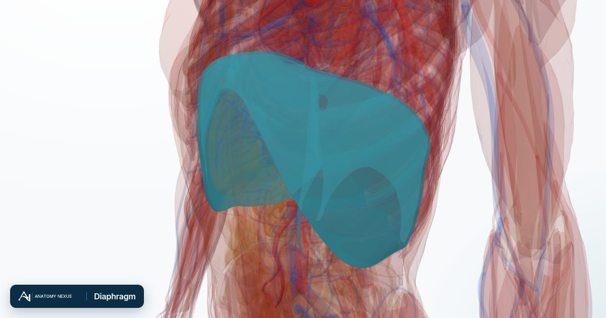
About
The dome-shaped sheet of muscle separating the chest from the abdomen.
Function. The main muscle of breathing: contracting flattens the dome, enlarging the chest and drawing air in. Openings carry the aorta, oesophagus and inferior vena cava.
Clinical note. Hiatus hernia lets the stomach slide through the oesophageal opening; the phrenic nerves that drive it arise from C3 to C5.
Structures in the atlas 1
The modelled pieces that make up the diaphragm in the 3D atlas. Each opens in the explorer.
The muscles in brief
Skeletal muscle makes up roughly 40% of body mass. Each muscle spans one or more joints, attaching to bone through tendons or flat aponeuroses; when its fibres shorten it pulls the bones together. Muscles work in groups: agonists produce a movement, antagonists control it, and stabilisers hold neighbouring joints still. The model includes the muscles of the limbs, trunk, neck, face, larynx and pharynx, and the diaphragm.