



Sensory organs
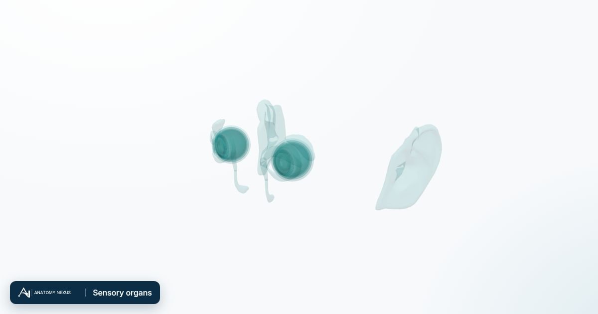
Eyes and ears with their internal structures.
Overview
The eye focuses light through the cornea and lens onto the retina, whose signals travel along the optic nerve to the brain. The model separates the cornea, sclera, choroid, iris, lens, vitreous body and retina, plus the lacrimal apparatus that produces and drains tears and the tarsal plates of the eyelids. The external ear collects sound towards the tympanic membrane; the middle and inner ear are not modelled in this release.
Functions
- Vision: focusing and detecting light
- Protecting and lubricating the eye (eyelids, tears)
- Hearing and balance (ear)
Clinical notes
- Refractive errors, cataract and glaucoma
- Retinal detachment and macular degeneration
- Conjunctivitis and blocked tear ducts
Organs & groups in this system
Eye
The eyeball and its parts: cornea, lens, iris, retina and the tissues that protect and move it.
Ear
The external ear gathers sound; the middle and inner ear (not modelled here) convert it to nerve signals and sense balance.
All structures 35
37 modelled pieces. Click a structure to open it in the atlas.
- Anterior chamber of left eyeball
- Anterior chamber of right eyeball
- External ear
- Left choroid · 2 pieces
- Left cornea
- Left corona ciliaris
- Left iris
- Left lacrimal canaliculus
- Left lacrimal gland
- Left lacrimal lake
- Left lacrimal sac
- Left lens
- Left nasolacrimal duct
- Left sclera
- Left vitreous body
- Optic part of left retina
- Optic part of right retina
- Right choroid · 2 pieces
- Right cornea
- Right corona ciliaris
- Right iris
- Right lacrimal canaliculus
- Right lacrimal gland
- Right lacrimal lake
- Right lacrimal sac
- Right lens
- Right nasolacrimal duct
- Right sclera
- Right vitreous body
- Suspensory ligament of left lens
- Suspensory ligament of right lens
- Tarsal plate of left lower eyelid
- Tarsal plate of left upper eyelid
- Tarsal plate of right lower eyelid
- Tarsal plate of right upper eyelid