



Anatomy · Sensory organs · Head
Eye anatomy
Also known as: eyes, eyeball, vision
The eyeball and its parts: cornea, lens, iris, retina and the tissues that protect and move it.
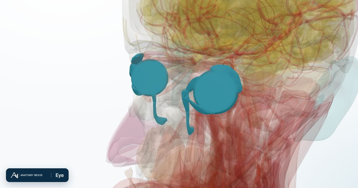
Overview page. A full anatomy article for the eye has not been written yet; below are the modelled structures, the system it belongs to and every clinical topic that mentions it.
Structures in the atlas 34
The modelled pieces that make up the eye in the 3D atlas. Each opens in the explorer.
- Anterior chamber of left eyeball
- Anterior chamber of right eyeball
- Left choroid · 2 pieces
- Left cornea
- Left corona ciliaris
- Left iris
- Left lacrimal canaliculus
- Left lacrimal gland
- Left lacrimal lake
- Left lacrimal sac
- Left lens
- Left nasolacrimal duct
- Left sclera
- Left vitreous body
- Optic part of left retina
- Optic part of right retina
- Right choroid · 2 pieces
- Right cornea
- Right corona ciliaris
- Right iris
- Right lacrimal canaliculus
- Right lacrimal gland
- Right lacrimal lake
- Right lacrimal sac
- Right lens
- Right nasolacrimal duct
- Right sclera
- Right vitreous body
- Suspensory ligament of left lens
- Suspensory ligament of right lens
- Tarsal plate of left lower eyelid
- Tarsal plate of left upper eyelid
- Tarsal plate of right lower eyelid
- Tarsal plate of right upper eyelid
The sensory organs in brief
The eye focuses light through the cornea and lens onto the retina, whose signals travel along the optic nerve to the brain. The model separates the cornea, sclera, choroid, iris, lens, vitreous body and retina, plus the lacrimal apparatus that produces and drains tears and the tarsal plates of the eyelids. The external ear collects sound towards the tympanic membrane; the middle and inner ear are not modelled in this release.