



Urinary catheterisation
Also known as: urinary catheter, Foley catheter, indwelling catheter, intermittent self-catheterisation, suprapubic catheter
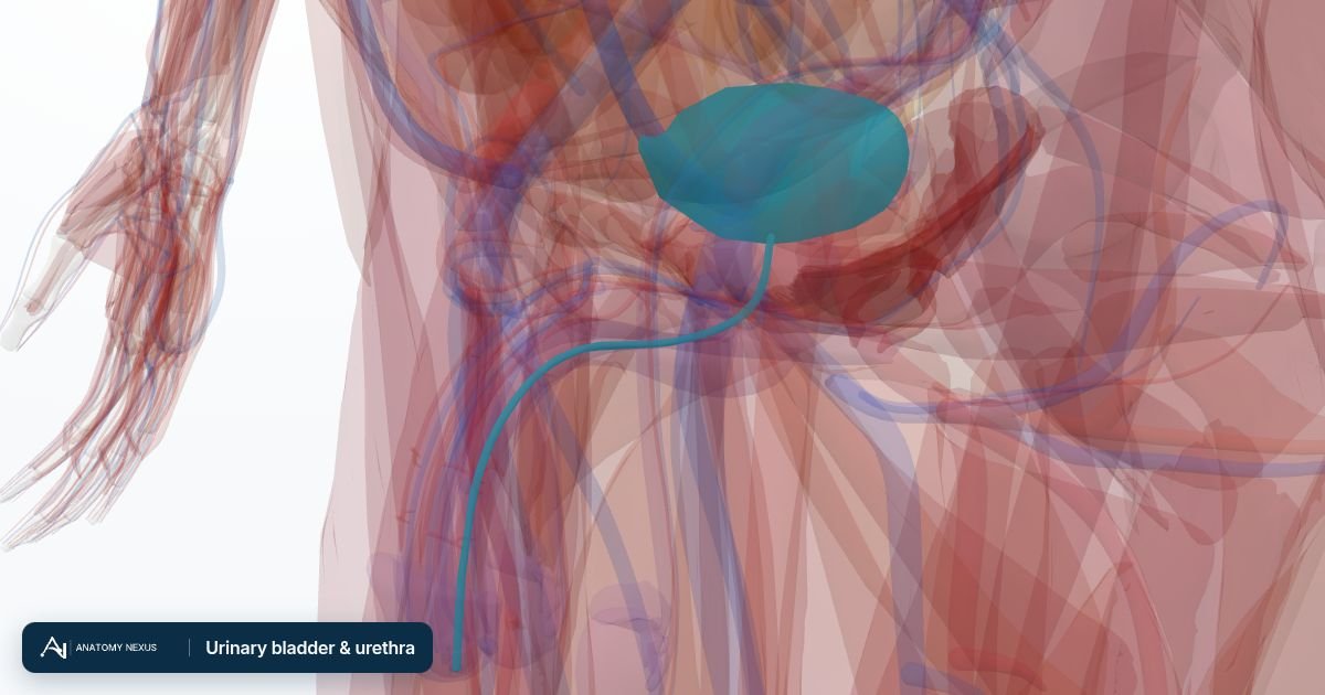
A soft tube passed along the urethra into the bladder to drain urine, held in place by a small balloon inflated inside the bladder. Used for hours during surgery, days on a ward, or long term when the bladder cannot empty. A suprapubic catheter enters through the lower abdominal wall instead; intermittent catheterisation passes a tube several times a day and removes it each time.
What it is
A soft tube passed along the urethra into the bladder to drain urine, held in place by a small balloon inflated inside the bladder. Used for hours during surgery, days on a ward, or long term when the bladder cannot empty. A suprapubic catheter enters through the lower abdominal wall instead; intermittent catheterisation passes a tube several times a day and removes it each time.
Why it is done
- Acute urinary retention (usually from an enlarged prostate) or chronic retention with kidney damage
- Measuring urine output hour by hour in critical illness and major surgery
- Neurological bladder problems (spinal cord injury, multiple sclerosis), often by self-catheterisation
- Bladder irrigation after prostate or bladder surgery, or with clots
- Incontinence with skin breakdown when other measures fail (last resort)
Before the procedure
- Explanation and consent; checking for latex allergy and previous difficult catheters
- Sterile technique: hand hygiene, gloves, cleaning the urethral opening
- Anaesthetic lubricant gel instilled into the urethra and left for a few minutes
Step by step
- The catheter (usually 12-16 Fr) is advanced gently along the urethra; in men the penis is held upright to straighten the first bend, then lowered to pass the prostate
- Urine flows once the tip is in the bladder; the catheter is advanced further before the balloon is inflated with 10 mL of sterile water
- It is drawn back until the balloon rests at the bladder neck and connected to a drainage bag or valve
- The volume drained is recorded; very large volumes are drained in stages if chronic retention is suspected
- The catheter is secured to the thigh to prevent traction
Afterwards
- Daily cleaning of the meatus, keeping the bag below bladder level and changing bags on schedule
- Removal (trial without catheter) as soon as it is no longer needed, usually within days
- Long-term catheters are changed every 4-12 weeks; bacteria in the urine are expected and not treated unless there are symptoms
Recovery
Slight stinging on first passing urine after removal is normal; a failed trial without catheter leads to re-catheterisation and urology review.
Risks
- Urinary tract infection, rising with each day in place, and catheter-related sepsis
- Urethral trauma, bleeding and false passage, especially in men with strictures or enlarged prostates
- Blockage by debris or clots; bypassing; bladder spasm
- Long term: strictures, stones, bladder cancer risk after many years
Alternatives
- Bladder scan and watchful measures for mild retention
- Alpha-blocker tablets before a trial without catheter in prostatic retention
- Intermittent self-catheterisation or suprapubic catheter for long-term needs
- Sheath (condom) drainage or pads for incontinence